. 2001 Dec 17;20(24):7313-22.
doi: 10.1093/emboj/20.24.7313.

The RecOR proteins modulate RecA protein function at 5' ends of single-stranded DNA

Affiliations

The RecOR proteins modulate RecA protein function at 5' ends of single-stranded DNA

J M Bork et al. EMBO J. .

Abstract

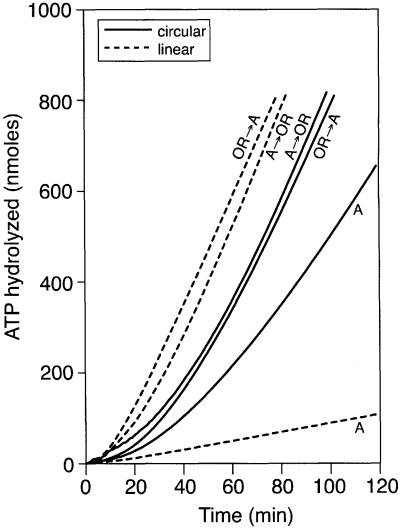
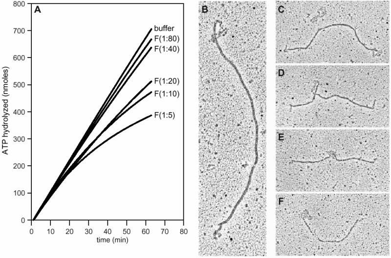
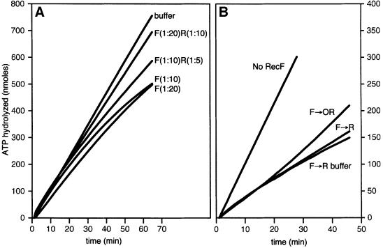
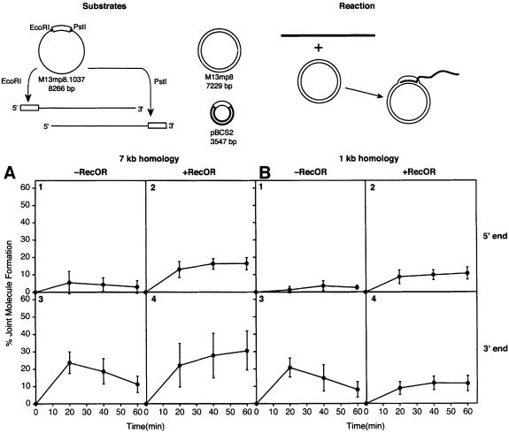
The Escherichia coli RecF, RecO and RecR pro teins have previously been implicated in bacterial recombinational DNA repair at DNA gaps. The RecOR-facilitated binding of RecA protein to single-stranded DNA (ssDNA) that is bound by single-stranded DNA-binding protein (SSB) is much faster if the ssDNA is linear, suggesting that a DNA end (rather than a gap) facilitates binding. In addition, the RecOR complex facilitates RecA protein-mediated D-loop formation at the 5' ends of linear ssDNAs. RecR protein remains associated with the RecA filament and its continued presence is required to prevent filament disassembly. RecF protein competes with RecO protein for RecR protein association and its addition destabilizes RecAOR filaments. An enhanced function of the RecO and RecR proteins can thus be seen in vitro at the 5' ends of linear ssDNA that is not as evident in DNA gaps. This function is countered by the RecF/RecO competition for association with the RecR protein.


Figures

None
Fig. 1. The RecOR proteins promote RecA filament assembly more efficiently on SSB-coated linear ssDNA than on SSB-coated circular ssDNA. The ssDNA-dependent ATPase assay was used to monitor RecA binding to ssDNA. The SSB protein (0.3 µM) was pre-bound to either M13mp8.1037(+) circular ssDNA (3 µM) or M13mp8.1037(+)xPstI linear ssDNA (3 µM). After a 10 min pre-incubation at 37°C, ATP (3 mM) and either RecA protein (4 µM) or the RecO (0.03 µM) and RecR (0.15 µM) proteins were added, as indicated. After another 10 min, the RecO and RecR proteins were added to the RecA reactions and vice versa. The addition of RecA protein defines t = 0. The circular ssDNA reactions are indicated with solid lines and the linear ssDNA reactions are indicated with dashed lines.
None
Fig. 2. The RecR protein is associated with linear ssDNA coated with SSB. M13mp8.1037(+)xEcoRI (12 µM) was incubated with SSB (1.2 µM) in HEPES buffer and the PEP/PK regeneration system. The samples were cross-linked, dialyzed and spread by the cytochrome C method, as described in Materials and methods. (AD) Representative molecules are shown in the micrographs for the reaction in the absence (A and B) or presence (C and D) of the RecO (0.12 µM) and RecR (0.6 µM) proteins. (E and F) An immunoaffinity labeling procedure identified RecR protein associated with linear ssDNA bound by SSB in the presence of the RecO and RecR proteins. RecR antibodies were diluted 1000-fold and protein A–gold was diluted 20-fold. (G) The number of gold particles per molecule were counted on a sample similar to those in (E) and (F) except that the RecR antibody was diluted 500-fold. The average number of gold particles per molecule for this labeling was six RecR-labeled gold particles per molecule.
None
Fig. 3. The RecR protein remains associated with a RecA filament after its formation. The RecA protein (5 µM) was incubated with M13mp8.1037(+) xEcoRI (12 µM) in HEPES buffer with the PEP/PK regeneration system. The SSB protein (1.2 µM), RecO protein (0.2 µM) and RecR protein (0.6 µM) were added to the reaction after an initial 10 min incubation at 37°C. After 15 min, the reaction was stopped with ATPγS and diluted 60-fold before spreading on Alcian-activated grids (described in Materials and methods). An immunoaffinity labeling procedure identified RecR protein that is associated with the RecA filament, as seen in the micrographs, with a 5000-fold dilution of RecR antibody and 20-fold dilution of protein A–gold (arrows). One hundred and thirty-eight molecules were analyzed. (A) Judgements were made as to where the gold-labeled RecR protein was found along the molecule. Molecules were divided into quarters and the number of gold particles in each segment of each molecule was counted. The majority of gold particles were found to be located within one end segment of the molecules, which is oriented to the left on the micrographs and designated quarter A in the histogram. The only gold particles found in quarter D were on molecules that had gold particles associated at both ends. If a molecule did not have any gold particles located in an end segment, then defining the placement of the golds into either quarter B or C was arbitrary. (B) The histogram shows the number of gold particles per molecule, the average being 3.5 RecR-labeled gold particles per molecule.
None
Fig. 4. The RecOR proteins facilitate RecA binding to gDNA–SSB molecules. Electron microscopy allowed visualization of RecA filaments formed on circular gDNA in the presence or absence of the RecOR proteins in Tris buffer. Reactions were analyzed after 8 and 20 min of incubation. SSB (0.051 µM or one monomer per 10 nt of ssDNA) was pre-incubated with GD2089 (3 µM total nt) in the presence of the PC/CPK regeneration system, as described in Materials and methods. The pre-incubation was 5 min at 37°C, at which point RecA protein (3 µM) and ATP (3 mM) were added. In the absence of the RecOR proteins, samples were taken after 8 and 20 min. Otherwise, RecA was incubated with GD2089–SSB for 5 min, then RecO (0.03 µM) and RecR (0.15 µM) proteins were added. Samples were taken after 8 and 20 min. Reactions were stopped with ATPγS, diluted 10-fold and spread on BSA-activated grids, as described in Materials and methods. The numbers of molecules with full RecA filaments (A), partial RecA filaments (B), mixed RecA and SSB proteins (C) and no RecA protein bound (D) were counted and are shown as percentages of all the molecules counted. For the 8 min samples, n = 299 in the absence of RecOR and n = 249 in the presence of RecOR. For the 20 min samples, n = 209 in the absence of RecOR and n = 133 in the presence of RecOR.
None
Fig. 5. The RecF protein destabilizes linear ssDNA–RecAOR filaments. (A) The ssDNA-dependent ATPase assay was used to monitor the stability of RecA protein filaments on linear ssDNA. A stable RecA filament (3 µM) was formed on M13mp8.1037(+)xPstI (3 µM) in the presence of SSB (0.3 µM), RecO (0.03 µM) and RecR (0.15 µM) proteins. The reaction was carried out as described in Materials and methods. RecF protein concentrations are expressed as monomer per nucleotide of ssDNA. At t = 0, the stable linear ssDNA–RecAOR filament was challenged by the addition of increasing amounts of RecF protein: no RecF or RecF at 1:80, 1:40, 1:20, 1:10 and 1:5. (BF) EM allowed visualization of linear ssDNA–RecAOR filaments in the presence of the RecF protein. A stable RecA filament (5 µM) was formed on M13mp8.1037(+)xPstI (12 µM) in the presence of SSB (1.2 µM), RecO (0.12 µM) and RecR (0.6 µM) proteins with the PEP/PK regeneration system for 15 min at 37°C. The RecF protein (1.2 µM) was added at 1:10. After a 20 min incubation, reactions were stopped with ATPγS, diluted 60-fold and spread as described in Materials and methods.
None
Fig. 6. Competition between RecF and RecO proteins for RecR protein. The ssDNA-dependent ATPase assay was used to monitor the stability of RecA protein filaments on linear ssDNA. A stable RecA filament (3 µM) was formed on M13mp8.1037(+)xPstI (3 µM) in the presence of SSB (0.3 µM), RecO (0.03 µM) and RecR (0.15 µM) proteins. Reactions were carried out as described in Materials and methods. Challenging protein concentrations are expressed as monomer per nucleotide of ssDNA. (A) At t = 0, the linear ssDNA–RecAOR filament was challenged with either RecF protein alone (1:20) or (1:10) or with the RecFR proteins together (1:20; 1:10) or (1:10; 1:5). (B) The linear ssDNA–RecAOR filament was challenged with RecF protein (1:10) or RecF buffer (no RecF). At t = 0, RecR (1:20), RecOR (1:100; 1:20) or RecR buffer (no RecR) were added to the three reactions that were challenged with RecF protein.
None
Fig. 7. Effects of the RecO and RecR proteins on RecA-mediated DNA pairing. The DNA substrates and the reaction are illustrated at the top. M13mp8.1037(+) circular ssDNA was linearized with either EcoRI or PstI to place the 1037 nt insert (open rectangle) at the 5′ or 3′ end, respectively. The supercoiled M13mp8 dsDNA substrate has 7229 bp of homology to the 3′ end of M13mp8.1037(+)xEcoRI linear ssDNA and the 5′ end of M13mp8.1037(+)xPstI linear ssDNA. The supercoiled pBCS2 dsDNA substrate has 1034 bp of homology (open rectangle) to the 5′ end of M13mp8.1037(+)xEcoRI linear ssDNA and the 3′ end of M13mp8.1037(+)xPstI linear ssDNA. (A and B) Four reactions using dsDNA substrates with 7229 bp (M13mp8) and 1034 bp (pBCS2) of homology, respectively, with the linear ssDNA substrate. Within each set of four, reactions 1 and 2 show results with the homology located at the 5′ end of the linear ssDNA, while reactions 3 and 4 show results with the homology located at the 3′ end. In addition, reactions 1 and 3 lack the RecOR proteins, while reactions 2 and 4 contain them. For (A), n = 7, 7, 9 and 8 for reactions 1–4, respectively. For (B), n = 7, 7, 9 and 8 for reactions 1–4, respectively. Reactions were carried out as described in Materials and methods for the agarose gel assay.

References

    1. Beernink H.T.H. and Morrical,S.W. (1999) RMPs: recombination/replication mediator proteins. Trends Biochem. Sci., 24, 385–389. - PubMed
    1. Bleuit J.S., Xu,H., Ma,Y.J., Wang,T.S., Liu,J. and Morrical,S.W. (2001) Mediator proteins orchestrate enzyme–ssDNA assembly during T4 recombination-dependent DNA replication and repair. Proc. Natl Acad. Sci. USA, 98, 8298–8305. - PMC - PubMed
    1. Bork J.M., Cox,M.M. and Inman,R.B. (2001) RecA protein filaments disassemble in the 5′ to 3′ direction on single-stranded DNA. J. Biol. Chem., 276, in press. - PubMed
    1. Bradford M.M. (1976) A rapid and sensitive method for the quantitation of microgram quantities of protein utilizing the principle of protein–dye binding. Anal. Biochem., 72, 248–254. - PubMed
    1. Clark A.J. and Sandler,S.J. (1994) Homologous genetic recombination: the pieces begin to fall into place. Crit. Rev. Microbiol., 20, 125–142. - PubMed

Publication types