

Position-effect variegation in Drosophila: the modifier Su(var)3-7 is a modular DNA-binding protein
- PMID: 11743022
- PMCID: PMC1084161
- DOI: 10.1093/embo-reports/kve243
Position-effect variegation in Drosophila: the modifier Su(var)3-7 is a modular DNA-binding protein
Abstract
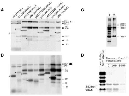
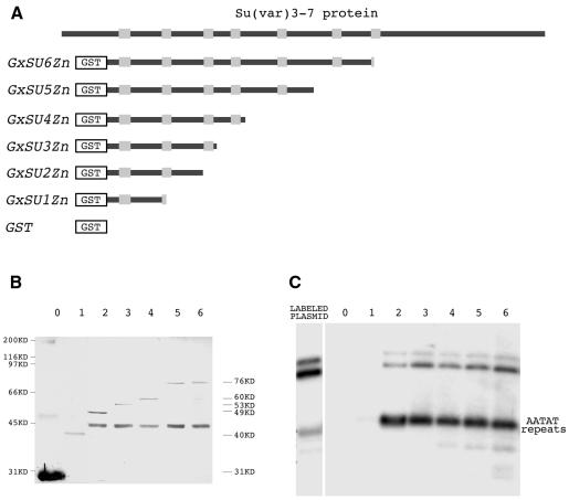
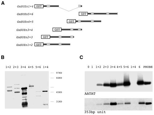
An increase in the dose of the Su(var)3-7 locus of Drosophila augments heterochromatin-promoted variegated silencing. The deduced protein sequence of Su(var)3-7 reveals seven widely spaced zinc fingers. We found that Su(var)3-7 has affinity for DNA in vitro and that the minimal protein sequence requirement for DNA binding is any module containing two zinc fingers and the interval between them. As Su(var)3-7 is a heterochromatin-associated protein, we tested its affinity for various satellite DNA sequences in vitro. The AATAT and 353-bp elements have the highest affinity. If affinity for satellite DNAs contributes to the presence of Su(var)3-7 in heterochromatin, a general affinity for DNA, or sequences yet to be determined, suggests a function in the genomic silencing of position-effect variegation: expansion of heterochromatin, whether continuous by spreading or discontinuous by pairing with sequence elements scattered through euchromatin, could use the affinity of Su(var)3-7 for DNA.
Figures
Similar articles
-
Conserved domains control heterochromatin localization and silencing properties of SU(VAR)3-7.Chromosoma. 2006 Apr;115(2):139-50. doi: 10.1007/s00412-005-0036-2. Epub 2006 Feb 7. Chromosoma. 2006. PMID: 16463146
-
Functional dissection of the Drosophila modifier of variegation Su(var)3-7.Development. 2002 Sep;129(17):3975-82. doi: 10.1242/dev.129.17.3975. Development. 2002. PMID: 12163401
-
Sumoylation of Drosophila SU(VAR)3-7 is required for its heterochromatic function.Nucleic Acids Res. 2010 Jul;38(13):4254-62. doi: 10.1093/nar/gkq168. Epub 2010 Mar 18. Nucleic Acids Res. 2010. PMID: 20299342 Free PMC article.
-
Histone modification and the control of heterochromatic gene silencing in Drosophila.Chromosome Res. 2006;14(4):377-92. doi: 10.1007/s10577-006-1066-1. Chromosome Res. 2006. PMID: 16821134 Review.
-
SU(VAR)3-9 is a conserved key function in heterochromatic gene silencing.Genetica. 2003 Mar;117(2-3):149-58. doi: 10.1023/a:1022923508198. Genetica. 2003. PMID: 12723694 Review.
Cited by
-
Isolation of Su(var)3-7 mutations by homologous recombination in Drosophila melanogaster.Genetics. 2002 Jul;161(3):1125-36. doi: 10.1093/genetics/161.3.1125. Genetics. 2002. PMID: 12136016 Free PMC article.
-
Modification of position-effect variegation by competition for binding to Drosophila satellites.EMBO Rep. 2002 Aug;3(8):747-52. doi: 10.1093/embo-reports/kvf155. Epub 2002 Jul 15. EMBO Rep. 2002. PMID: 12151333 Free PMC article.
-
The recently identified modifier of murine metastable epialleles, Rearranged L-Myc Fusion, is involved in maintaining epigenetic marks at CpG island shores and enhancers.BMC Biol. 2015 Mar 26;13:21. doi: 10.1186/s12915-015-0128-2. BMC Biol. 2015. PMID: 25857663 Free PMC article.
-
A transcription factor-based mechanism for mouse heterochromatin formation.Nat Struct Mol Biol. 2012 Oct;19(10):1023-30. doi: 10.1038/nsmb.2382. Epub 2012 Sep 16. Nat Struct Mol Biol. 2012. PMID: 22983563
-
Conserved domains control heterochromatin localization and silencing properties of SU(VAR)3-7.Chromosoma. 2006 Apr;115(2):139-50. doi: 10.1007/s00412-005-0036-2. Epub 2006 Feb 7. Chromosoma. 2006. PMID: 16463146
References
-
- Belyaeva E.S. and Zhimulev, I.F. (1991) Cytogenetic and molecular aspects of position-effect variegation in Drosophila melanogaster. III. Continuous and discontinuous compaction of chromosomal material as a result of position effect variegation. Chromosoma, 100, 453–466. - PubMed
-
- Cortés A., Huertas, D., Fanti, L., Pimpinelli, S., Marsellach, F.X., Pina, B. and Azorin, F. (1999) DDP1, a single-stranded nucleic acid-binding protein of Drosophila, associates with pericentric heterochromatin and is functionally homologous to the yeast Scp160p, which is involved in the control of cell ploidy. EMBO J., 18, 3820–3833. - PMC - PubMed
Publication types
MeSH terms
Substances
LinkOut - more resources
Full Text Sources
Molecular Biology Databases
Research Materials
