. 2002 Feb 22;277(8):6183-7.
doi: 10.1074/jbc.M110978200. Epub 2001 Dec 14.

Hypoxia-inducible factor-1-mediated expression of the 6-phosphofructo-2-kinase/fructose-2,6-bisphosphatase-3 (PFKFB3) gene. Its possible role in the Warburg effect

Affiliations

Hypoxia-inducible factor-1-mediated expression of the 6-phosphofructo-2-kinase/fructose-2,6-bisphosphatase-3 (PFKFB3) gene. Its possible role in the Warburg effect

Alexander Minchenko et al. J Biol Chem. .

Abstract

One of the key mediators of the hypoxic response in animal cells is the hypoxia-inducible transcription factor-1 (HIF-1) complex, in which the alpha-subunit is highly susceptible to oxygen-dependent degradation. The hypoxic response is manifested in many pathophysiological processes such as tumor growth and metastasis. During hypoxia, cells shift to a primarily glycolytic metabolic mode for their energetic needs. This is also manifested in the HIF-1-dependent up-regulation of many glycolytic genes. Paradoxically, tumor cells growing under conditions of normal oxygen tension also show elevated glycolytic rates that correlate with the increased expression of glycolytic enzymes and glucose transporters (the Warburg effect). A key regulator of glycolytic flux is the relatively recently discovered fructose-2,6-bisphosphate (F-2,6-P2), an allosteric activator of 6-phosphofructo-1-kinase (PFK-1). Steady state levels of F-2,6-P2 are maintained by the bifunctional enzyme PFK-2/F2,6-Bpase, which has both kinase and phosphatase activities. Herein, we show that one isozyme, PFKFB3, is highly induced by hypoxia and the hypoxia mimics cobalt and desferrioxamine. This induction could be replicated by the use of an inhibitor of the prolyl hydroxylase enzymes responsible for the von Hippel Lindau (VHL)-dependent destabilization and tagging of HIF-1 alpha. The absolute dependence of the PFKFB3 gene on HIF-1 was confirmed by its overexpression in VHL-deficient cells and by the lack of hypoxic induction in mouse embryonic fibroblasts conditionally nullizygous for HIF-1 alpha.


Figures

Fig. 1
Fig. 1. Response of the PFKFB3 gene to hypoxia, cobalt, and desferrioxamine
a, Hep-3B cells were maintained in normoxia (N), exposed to hypoxia (H), 0.5% O2, or incubated with 100 µm cobalt chloride (Co) or 130 µm desferrioxamine (D) for 6 h. Total RNA was extracted and analyzed for PFKFB3 and Glut-1 mRNA expression using RNase protection assays. b, Hep-3B cells where treated with desferroxamine, and total RNA was isolated at various times (Hrs) following the treatment and analyzed for Glut-1 (left) or PFKGFB3 (right) mRNA. c, responsiveness of the PFKFB3 gene to hypoxia (H) and desferrioxamine in wild-type (W.T.) and ρ° cells (25). In all figures, 18s represents 18 S ribosomal RNA.
Fig. 2
Fig. 2. Hypoxic response of the PFKFB3 gene in HIF-1α negative cells
a, HIF-1α negative mouse fibroblasts (−) and their HIF-1α positive (+) controls (14, 34) where exposed to hypoxia for 6 h, and the total RNA was analyzed for PFKFB3 and Glut-1 mRNA by RNase protection assays. b, electrophoretic gel shift assay of nuclear extracts from HIF-1α (+) and HIF-1α (−) cells exposed to normoxia (N) or hypoxia (H) using a probe (P) that contains the erythropoietin HRE. C represents constitutive bands. The rightmost panel shows a supershift assay in hypoxic HIF-1α (+) cells utilizing anti-HIF-1α antibodies.
Fig. 3
Fig. 3. Effect of dimethyloxalylglycine on PFKFB3 mRNA expression
a, confluent monolayers of hepatoma (Hep3B) or retinal pigment epithelial (RPE) cells where maintained in normoxia (N) or exposed to 1 mm dimethyloxalylglycine (I) or hypoxia (H) for 6 h. Total RNA was analyzed for PFKFB3 and Glut-1 expression by RNase protection assay. b, Western blot of Hep-3B cells treated as above and analyzed for HIF-1α expression utilizing anti-HIF-1α monoclonal antibody.
Fig. 4
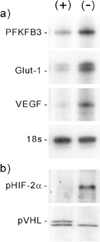
Fig. 4. Expression of the PFKFB3 gene in pVHL-deficient cells
a, total RNA from normoxic VHL deficient (−) renal carcinoma 786-0 cells and their VHL positive (+) controls where analyzed for PFKFB3, Glut-1, and VEGF mRNA expression by RNase protection assays. b, whole cell extracts from VHL (+) and VHL (−) were analyzed by Western blots using anti-HIF-2α and anti-VHL antibodies.

References

    1. Kawaguchi T, Veech RL, Uyeda K. J. Biol. Chem. 2001;276:28554–28561. - PubMed
    1. Hue L, Rousseau GG. Adv. Enzyme Regul. 1993;33:97–110. - PubMed
    1. Okar DA, Lange AJ. Biofactors. 1999;10:1–14. - PubMed
    1. Pilkis SJ, Claus TH, Kurland IJ, Lange AJ. Annu. Rev. Biochem. 1995;64:799–835. - PubMed
    1. Chesney J, Mitchell R, Benigni F, Bacher M, Spiegel L, Al-Abed Y, Han JH, Metz C, Bucala R. Proc. Natl. Acad. Sci. U. S. A. 1999;96:3047–3052. - PMC - PubMed

Publication types

MeSH terms

LinkOut - more resources