

Plasmidic versus insertional cloning of heterologous genes in Mycobacterium bovis BCG: impact on in vivo antigen persistence and immune responses
- PMID: 11748196
- PMCID: PMC127622
- DOI: 10.1128/IAI.70.1.303-314.2002
Plasmidic versus insertional cloning of heterologous genes in Mycobacterium bovis BCG: impact on in vivo antigen persistence and immune responses
Abstract
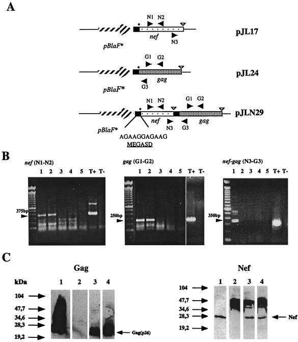
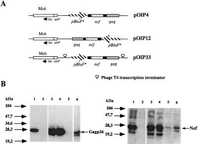
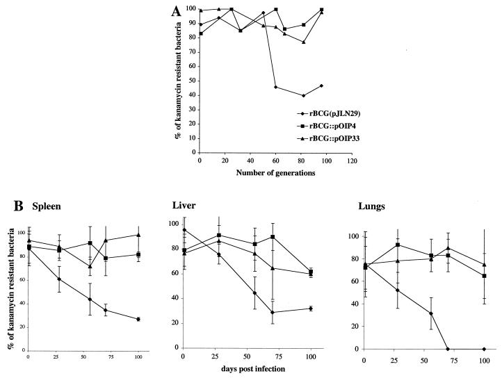
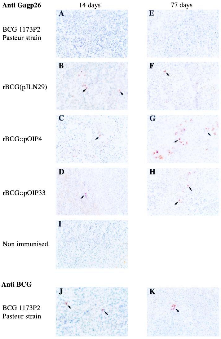
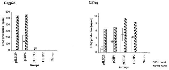
Bivalent recombinant strains of Mycobacterium bovis BCG (rBCG) expressing the early regulatory nef and the structural gag(p26) genes from the simian immunodeficiency virus (SIV) SIVmac251 were engineered so that both genes were cotranscribed from a synthetic operon. The expression cassette was cloned into a multicopy-replicating vector, and the expression levels of both nef and gag in the bivalent rBCG(nef-gag) strain were found to be comparable to those of monovalent rBCG(nef) or rBCG(gag) strains. However, extrachromosomal cloning of the nef-gag operon into a replicative plasmid resulted in strains of low genetic stability that rapidly lost the plasmid in vivo. Thus, the nef-gag operon was inserted site specifically into the BCG chromosome by means of mycobacteriophage Ms6-derived vectors. The resulting integrative rBCG(nef-gag) strains showed very high genetic stability both in vitro and in vivo. The in vivo expression of the heterologous genes was much longer lived when the expression cassette was inserted into the BCG chromosome. In one of the strains obtained, integrative cloning did not reduce the expression levels of the genes even though a single copy was present. Accordingly, this strain induced cellular immune responses of the same magnitude as that of the replicative rBCG strain containing several copies of the genes.
Figures
Similar articles
-
Recombinant Mycobacterium bovis BCG producing the N-terminal half of SIVmac251 Env antigen induces neutralizing antibodies and cytotoxic T lymphocyte responses in mice and guinea pigs.AIDS Res Hum Retroviruses. 1997 Dec 10;13(18):1573-81. doi: 10.1089/aid.1997.13.1573. AIDS Res Hum Retroviruses. 1997. PMID: 9430249
-
Oral immunization with recombinant Mycobacterium bovis BCG simian immunodeficiency virus nef induces local and systemic cytotoxic T-lymphocyte responses in mice.J Virol. 1997 Mar;71(3):2303-9. doi: 10.1128/JVI.71.3.2303-2309.1997. J Virol. 1997. PMID: 9032366 Free PMC article.
-
A cocktail of Mycobacterium bovis BCG recombinants expressing the SIV Nef, Env, and Gag antigens induces antibody and cytotoxic responses in mice vaccinated by different mucosal routes.AIDS Res Hum Retroviruses. 1998 Dec 20;14(18):1625-33. doi: 10.1089/aid.1998.14.1625. AIDS Res Hum Retroviruses. 1998. PMID: 9870315
-
Intradermal and oral immunization with recombinant Mycobacterium bovis BCG expressing the simian immunodeficiency virus Gag protein induces long-lasting, antigen-specific immune responses in guinea pigs.Clin Immunol. 2006 Apr;119(1):67-78. doi: 10.1016/j.clim.2005.11.005. Epub 2006 Jan 4. Clin Immunol. 2006. PMID: 16386958
-
Towards new mycobacterial vaccines.Dev Biol Stand. 1994;82:171-8. Dev Biol Stand. 1994. PMID: 7958472 Review.
Cited by
-
Biosynthesis and translocation of unsulfated acyltrehaloses in Mycobacterium tuberculosis.J Biol Chem. 2014 Oct 3;289(40):27952-65. doi: 10.1074/jbc.M114.581199. Epub 2014 Aug 14. J Biol Chem. 2014. PMID: 25124040 Free PMC article.
-
Optimisation of bioluminescent reporters for use with mycobacteria.PLoS One. 2010 May 24;5(5):e10777. doi: 10.1371/journal.pone.0010777. PLoS One. 2010. PMID: 20520722 Free PMC article.
-
MTBVAC-Based TB-HIV Vaccine Is Safe, Elicits HIV-T Cell Responses, and Protects against Mycobacterium tuberculosis in Mice.Mol Ther Methods Clin Dev. 2019 Feb 7;13:253-264. doi: 10.1016/j.omtm.2019.01.014. eCollection 2019 Jun 14. Mol Ther Methods Clin Dev. 2019. PMID: 30859110 Free PMC article.
-
Role of succinyl substituents in the mannose-capping of lipoarabinomannan and control of inflammation in Mycobacterium tuberculosis infection.PLoS Pathog. 2023 Sep 5;19(9):e1011636. doi: 10.1371/journal.ppat.1011636. eCollection 2023 Sep. PLoS Pathog. 2023. PMID: 37669276 Free PMC article.
-
Expression of Exogenous Antigens in the Mycobacterium bovis BCG Vaccine via Non-genetic Surface Decoration with the Avidin-biotin System.J Vis Exp. 2018 Jan 31;(131):56421. doi: 10.3791/56421. J Vis Exp. 2018. PMID: 29443102 Free PMC article.
References
-
- Abdelhak, S., H. Louzir, J. Timm, L. Blel, Z. Benlasfar, M. Lagranderie, M. Gheorghiu, K. Dellagi, and B. Gicquel. 1995. Recombinant BCG expressing the Leishmania surface antigen Gp63 induces protective immunity against Leishmania major infection in BALB/c mice. Microbiology 141: 1585–1592. - PubMed
-
- Aldovini, A., and R. A. Young. 1991. Humoral and cell-mediated immune responses to live recombinant BCG-HIV vaccines. Nature 351: 479–482. - PubMed
-
- Barker, L. P., D. M. Brooks, and P. L. C. Small. 1998. The identification of Mycobacterium marinum genes differentially expressed in macrophage phagosomes using promoter fusions to green fluorescent protein. Mol. Microbiol. 29: 1167–1177. - PubMed
-
- Berthet, F. X., M. Lagranderie, P. Gounon, C. Laurent-Winter, D. Ensergueix, P. Chavarot, F. Thouron, E. Maranghi, V. Pelicic, D. Portnoi, G. Marchal, and B. Gicquel. 1998. Attenuation of virulence by disruption of the Mycobacterium tuberculosis erp gene. Science 282: 759–762. - PubMed
Publication types
MeSH terms
Substances
LinkOut - more resources
Full Text Sources
Other Literature Sources
