. 2002 Jan;70(1):368-79.
doi: 10.1128/IAI.70.1.368-379.2002.

Genomic approach for analysis of surface proteins in Chlamydia pneumoniae

Affiliations

Genomic approach for analysis of surface proteins in Chlamydia pneumoniae

Silvia Montigiani et al. Infect Immun. 2002 Jan.

Abstract

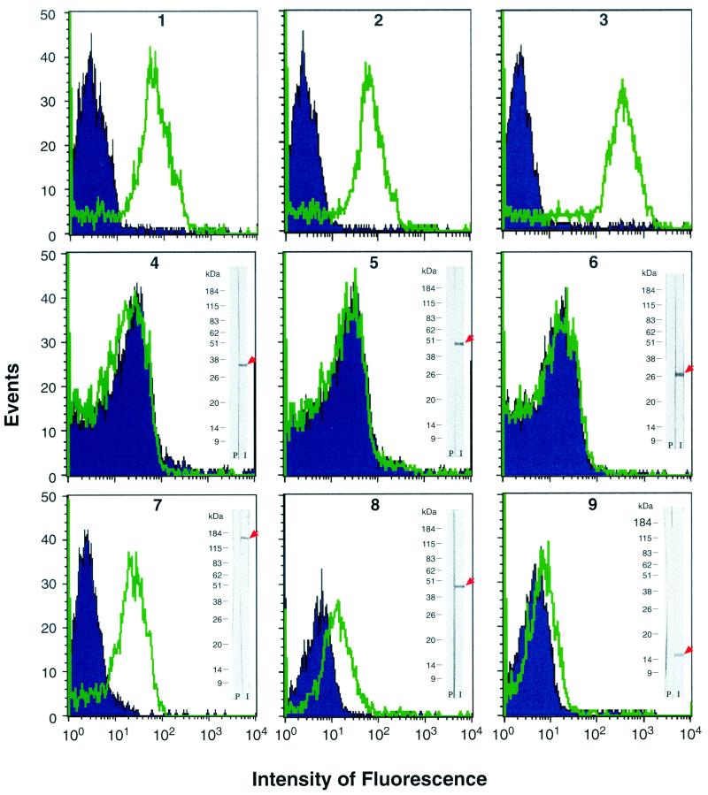
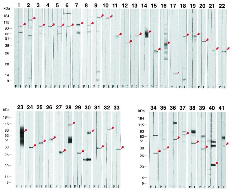
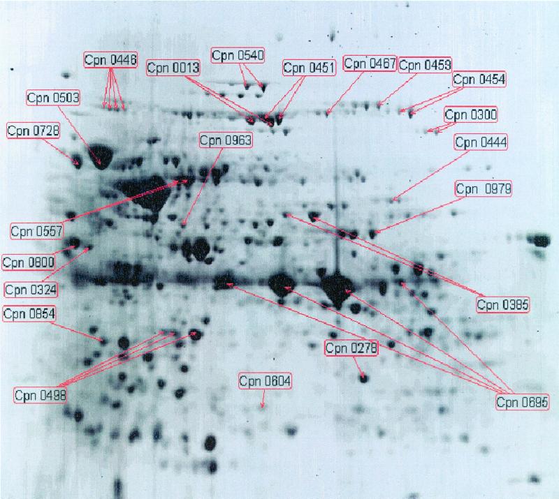
Chlamydia pneumoniae, a human pathogen causing respiratory infections and probably contributing to the development of atherosclerosis and heart disease, is an obligate intracellular parasite which for replication needs to productively interact with and enter human cells. Because of the intrinsic difficulty in working with C. pneumoniae and in the absence of reliable tools for its genetic manipulation, the molecular definition of the chlamydial cell surface is still limited, thus leaving the mechanisms of chlamydial entry largely unknown. In an effort to define the surface protein organization of C. pneumoniae, we have adopted a combined genomic-proteomic approach based on (i) in silico prediction from the available genome sequences of peripherally located proteins, (ii) heterologous expression and purification of selected proteins, (iii) production of mouse immune sera against the recombinant proteins to be used in Western blotting and fluorescence-activated cell sorter (FACS) analyses for the identification of surface antigens, and (iv) mass spectrometry analysis of two-dimensional electrophoresis (2DE) maps of chlamydial protein extracts to confirm the presence of the FACS-positive antigens in the chlamydial cell. Of the 53 FACS-positive sera, 41 recognized a protein species with the expected size on Western blots, and 28 of the 53 antigens shown to be surface-exposed by FACS were identified on 2DE maps of elementary-body extracts. This work represents the first systematic attempt to define surface protein organization in C. pneumoniae.


Figures

FIG. 1.
FIG. 1.
FACS analysis of antibody binding to whole C. pneumoniae EBs. Blue histograms (event counts versus fluorescence channels) are the FACS output for EBs stained with background control antibodies. Green histograms are the FACS output of EBs stained with antigen-specific antibodies. Positive controls (panels 1 to 3) were as follows: panel 1, anti-C. pneumoniae LPS monoclonal antibody, with an irrelevant monoclonal antibody (SEAM-3) specific for the type B meningococcal capsule as background control; panel 2, C. pneumoniae-specific anti-MOMP monoclonal antibody (Argene Biosoft), with an irrelevant fluorescein isothiocyanate-conjugated anti-immunoglobulin G2a monoclonal antibody as background control; panel 3, mouse hyperimmune serum against whole EBs, with the corresponding preimmune mouse serum as background control. Negative controls (panels 4 to 6), with mouse anti-GST serum as background control, were as follows: panel 4, mouse serum against 6482-GST fusion protein (PID accession no. 4376582; predicted as a cytoplasmic 36.8-kDa protein); panel 5, mouse serum against 6732-GST fusion protein (PID accession no. 4376732; predicted as a cytoplasmic 43.5-kDa protein); panel 6, mouse serum against 6881-GST fusion protein (PID accession no. 4376881; predicted as a cytoplasmic 26.0-kDa protein). Examples of FACS-positive sera (panels 7 to 9), with mouse anti-GST serum as background control, were as follows: panel 7, antiserum to 7287-GST antigen (Pmp-21); panel 8, antiserum to 6602-GST antigen (LrcE); panel 9, antiserum to 6577-GST antigen (annotated as an OmpH-like OMP). Western blotting data obtained from total EB proteins stained with the same antiserum used for the FACS assays are also shown.
FIG. 2.
FIG. 2.
Western blot analysis of total protein extracts from C. pneumoniae EBs, performed using mouse immune sera against recombinant antigens. For antigen identification, refer to Table 1. The panel identification numbers correspond to the numbers reported in the WB analysis column of Table 1. In each panel, the strip on the right shows the results obtained with the antigen-specific immune serum (I), and the strip on the left shows the results obtained with the corresponding preimmune serum (P). Panels 1 to 33 are data that were scored as “consistent” in Table 1, and panels 34 to 41 show results that were scored as “partially consistent” in Table 1.
FIG. 3.
FIG. 3.
2DE map of proteins from purified EBs of C. pneumoniae FB/96. The protein spots marked with the gene ID (see Table 2) correspond to FACS-positive antigens identified by MALDI-TOF analysis.

References

    1. Bavoil, P. M., R. C. Hsia, and D. Ojcius. 2000. Closing in on Chlamydia and its intracellular bag of tricks. Microbiology 146: 2723–2731. - PubMed
    1. Bavoil, P. M., and R. C Hsia. 1998. Type III secretion in Chlamydia: a case of deja vu? Mol. Microbiol. 28: 860–862. - PubMed
    1. Bjellqvist, B., G. J. Hughes, C. Pasquali, N. Paquet, F. Ravier, J. C. Sanchez, S. Frutiger, and D. Hochstrasser. 1993. The focusing positions of poly-peptides in immobilized pH gradients can be predicted from their amino acid sequences. Electrophoresis 14: 1023–1031. - PubMed
    1. Cheng, L. W., and O. Schneewind. 2000. Yersinia enterocolitica type A, an intracellular regulator of the type III machinery, is required for specific targeting of YopE, YopH, YopM, and YopN into the cytosol of eukaryotic cells. J. Bacteriol. 182: 3183–3190. - PMC - PubMed
    1. Chevallet, M., V. Santoni, A. Poinas, D. Rouquie, A. Fuchs, S. Kieffer, M. Rossignol, J. Lunardi, J. Garin, and T. Rabilloud. 1998. New zwitterionic detergents improve the analysis of membrane proteins by two-dimensional electrophoresis. Electrophoresis 19: 1901–1909. - PubMed

Publication types

MeSH terms

Substances