

Visualization of Proteus mirabilis within the matrix of urease-induced bladder stones during experimental urinary tract infection
- PMID: 11748205
- PMCID: PMC127628
- DOI: 10.1128/IAI.70.1.389-394.2002
Visualization of Proteus mirabilis within the matrix of urease-induced bladder stones during experimental urinary tract infection
Abstract
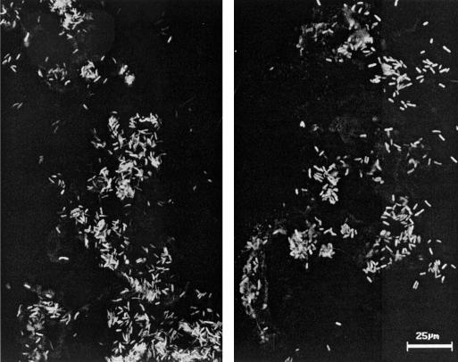
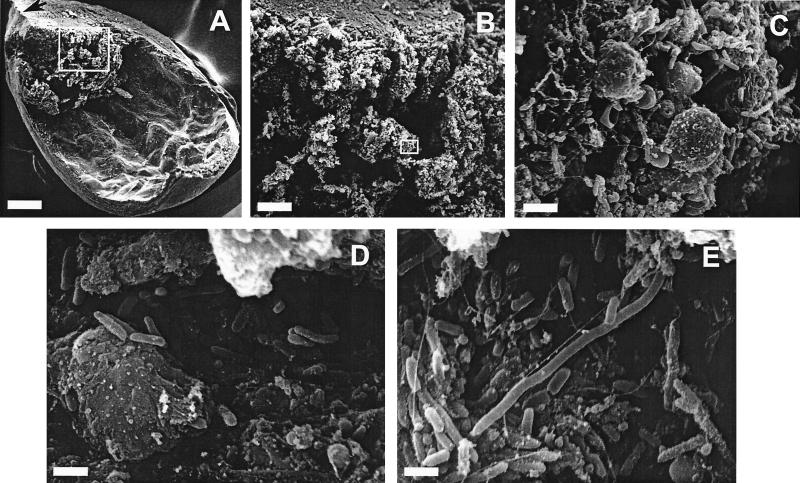
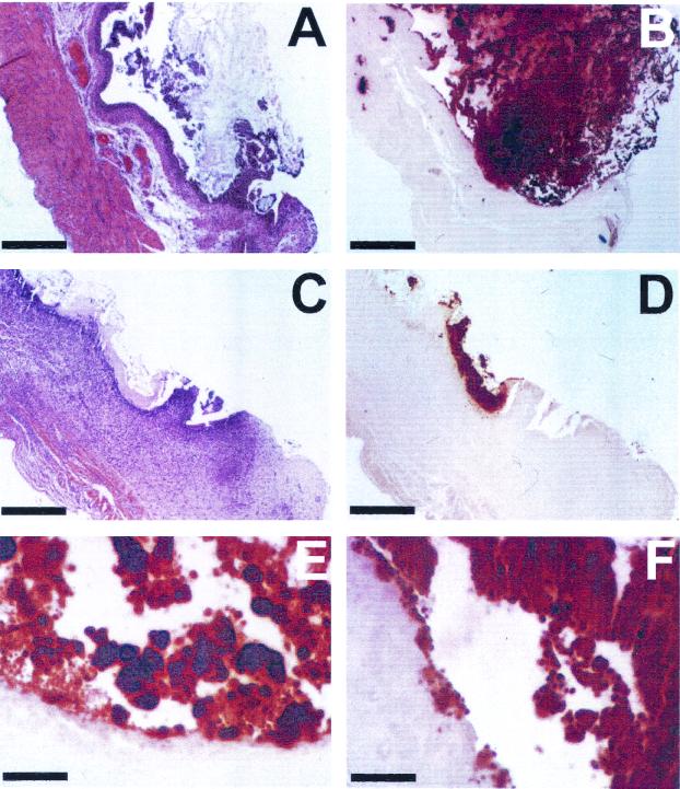
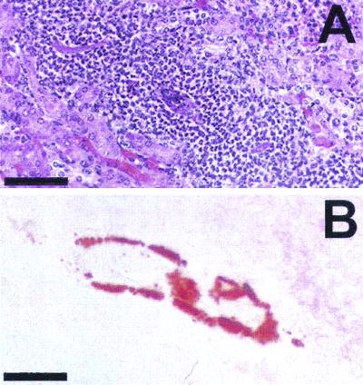
The virulence of a urease-negative mutant of uropathogenic Proteus mirabilis and its wild-type parent strain was assessed by using a CBA mouse model of catheterized urinary tract infection. Overall, catheterized mice were significantly more susceptible than uncatheterized mice to infection by wild-type P. mirabilis. At a high inoculum, the urease-negative mutant successfully colonized bladders of catheterized mice but did not cause urolithiasis and was still severely attenuated in its ability to ascend to kidneys. Using confocal laser scanning microscopy and scanning electron microscopy, we demonstrated the presence of P. mirabilis within the urease-induced stone matrix. Alizarin red S staining was used to detect calcium-containing deposits in bladder and kidney tissues of P. mirabilis-infected mice.
Figures
References
-
- Cohen, T. D., and G. M. Preminger. 1996. Struvite calculi. Semin. Nephrol. 16: 425–434. - PubMed
-
- Coker, C., C. A. Poore, X. Li, and H. L. T. Mobley. 2000. Pathogenesis of Proteus mirabilis urinary tract infection. Microbes Infect. 2: 1497–1505. - PubMed
-
- Cormack, B. P., R. H. Valdivia, and S. Falkow. 1996. FACS-optimized mutants of the green fluorescent protein (GFP). Gene 173: 33–38. - PubMed
-
- Fairley, K. F., N. E. Carson, R. C. Gutch, P. Leighton, A. D. Grounds, E. C. Laird, P. H. G. McCallum, R. L. Sleeman, and C. M. O’Keefe. 1971. Site of infection in acute urinary-tract infection in general practice. Lancet ii: 615–618. - PubMed
-
- Gilmore, S. K., S. W. Whitson, and D. E. Bowers, Jr. 1986. A simple method using alizarin red S for the detection of calcium in epoxy resin embedded tissue. Stain Technol. 61: 89–92. - PubMed
Publication types
MeSH terms
Substances
Grants and funding
LinkOut - more resources
Full Text Sources
