. 2001 Dec;108(12):1759-70.
doi: 10.1172/JCI12617.

Endothelial transcytosis of myeloperoxidase confers specificity to vascular ECM proteins as targets of tyrosine nitration

Affiliations

Endothelial transcytosis of myeloperoxidase confers specificity to vascular ECM proteins as targets of tyrosine nitration

S Baldus et al. J Clin Invest. 2001 Dec.

Abstract

Nitrotyrosine formation is a hallmark of vascular inflammation, with polymorphonuclear neutrophil-derived (PMN-derived) and monocyte-derived myeloperoxidase (MPO) being shown to catalyze this posttranslational protein modification via oxidation of nitrite (NO(2)(-)) to nitrogen dioxide (NO(2)(*)). Herein, we show that MPO concentrates in the subendothelial matrix of vascular tissues by a transcytotic mechanism and serves as a catalyst of ECM protein tyrosine nitration. Purified MPO and MPO released by intraluminal degranulation of activated human PMNs avidly bound to aortic endothelial cell glycosaminoglycans in both cell monolayer and isolated vessel models. Cell-bound MPO rapidly transcytosed intact endothelium and colocalized abluminally with the ECM protein fibronectin. In the presence of the substrates hydrogen peroxide (H(2)O(2)) and NO(2)(-), cell and vessel wall-associated MPO catalyzed nitration of ECM protein tyrosine residues, with fibronectin identified as a major target protein. Both heparin and the low-molecular weight heparin enoxaparin significantly inhibited MPO binding and protein nitrotyrosine (NO(2)Tyr) formation in both cultured endothelial cells and rat aortic tissues. MPO(-/-) mice treated with intraperitoneal zymosan had lower hepatic NO(2)Tyr/tyrosine ratios than did zymosan-treated wild-type mice. These data indicate that MPO significantly contributes to NO(2)Tyr formation in vivo. Moreover, transcytosis of MPO, occurring independently of leukocyte emigration, confers specificity to nitration of vascular matrix proteins.


Figures

Figure 1
Figure 1
Immunohistochemical detection of aortic endothelial distribution of MPO following activation of PMNs. Isolated human PMNs (8 × 10⁵) were instilled into the lumen of an excised rat aorta immediately after activation with 50 ng·ml–1 TPA. After 4 hours, aortic segments were frozen, sectioned, fixed with 4% paraformaldehyde, permeabilized using 0.1% Triton X-100, and analyzed by immunofluorescence. Tissue sections were stained for MPO (green) using rabbit anti-MPO and nuclei (blue) were counterstained with DAPI. (a) Untreated vessel; (b) vessel treated with TPA alone; (c) unstimulated PMNs; (d and e) stimulated PMNs. Colocalization of MPO with anti-vWF immunoreactivity confirmed MPO binding and uptake by endothelial cells (f). L, vessel lumen; M, media. Arrows indicate endothelial cells. ×100 (ad, f); ×50 (e).
Figure 2
Figure 2
Characterization of the binding and uptake of MPO by cultured endothelial cells. (a) Characterization of cell MPO association. BAECs were incubated with increasing concentrations of MPO (2–13 nM) for 2 hours at 37°C and harvested by scraping (total) or after exposure to trypsin (trypsin-resistant). Cell-associated MPO activity was about 10% of total MPO activity added (not shown). (b) The influence of temperature on cellular MPO binding and transport. BAECs exposed to MPO (13 nM) at 37°C and 4°C were harvested at different time points and MPO activity was determined. P < 0.05 for MPO activity in total cell lysates at 37°C versus 4°C; *P < 0.05 for MPO activity in trypsin-resistant compartments at 37°C versus 4°C. (c) The influence of glycosaminoglycans on cell MPO binding. BAECs were pretreated with chondroitin sulfate (Chondr, 150 μg·ml–1), heparin (Hep, 150 μg·ml–1), or the low–molecular weight heparin enoxaparin (Enox, 150 μg·ml–1) for 45 minutes, washed with HBSS, and then exposed to MPO (13 nM) at 37°C. After washing again, cell-associated MPO was assessed by enzyme activity analysis and cellular MPO protein content by immunoblotting. “MPO hc ” denotes the immunoreactivity of the heavy chain (59 kDa) of MPO. (d) The effect of endoglycosidases on MPO binding. BAECs were pretreated with heparitinase, heparinase, and chondroitinase (all 8 mU·ml–1) for 2 hours at 37°C, washed, and exposed to MPO (13 nM) at 4°C for 2 hours. Cell-associated MPO enzyme activity was then determined. *P < 0.05 for MPO alone versus pretreatment with heparin and enoxaparin (c) and versus heparitinase and heparinase pretreatment (d). (e) Binding competition analysis of MPO and xanthine oxidase (XO). BAECs were incubated with MPO (1 μg·ml–1) and increasing concentrations of XO (0–100 μg·ml–1, equivalent to 0–100 mU·ml–1) for 2 hours at 37°C. Cells were harvested as above and MPO enzyme activity determined. Values represent mean ± SD.
Figure 3
Figure 3
Transcytosis of MPO by endothelial cells. (a) Intracellular localization and accumulation of MPO at the ECM. Cultured BAECs were grown to confluence and exposed to MPO (13 nM) for 2 hours. Cells were processed as described in Methods and incubated with mouse monoclonal anti–α-tubulin (red) and rabbit polyclonal anti-MPO (green). Laser confocal microscopy revealed intracellular MPO immunoreactivity within 2 minutes after MPO exposure and a high degree of MPO deposition at the subcellular matrix. Time = 0 minutes represents BAECs not exposed to MPO but stained with anti-MPO. ×63. (b) Colocalization of MPO and fibronectin (FN). BAECs treated as described for a were incubated with mouse monoclonal anti-fibronectin (red) and rabbit polyclonal anti-MPO (green). The nuclei (blue) were counterstained with DAPI. For assessing codistribution of MPO and fibronectin, images were merged (yellow). The control panel (Ctr) shows MPO-untreated cells stained with anti-MPO. The lower panel represents side-on views. ×63 (control, ×50). (c) Barrier function of endothelial cell monolayers exposed to MPO. BAECs were seeded on Transwell filters and exposed to increasing concentrations of MPO (13–130 nM) in the presence of TRITC-labeled dextran (4,400 Da) for 2 hours. MPO activity and TRITC fluorescence were assessed in the basolateral chamber. Treatment of the cells with H2O2 (100 μM) served as a control for increased permeability of TRITC-dextran. *P > 0.05 versus TRITC-labeled dextran alone. RFU, relative fluorescence units.
Figure 4
Figure 4
MPO-dependent tyrosine nitration of ECM fibronectin. (a) NO2Tyr formation in cultured endothelial cell ECM proteins. Confluent BAEC monolayers were exposed to MPO (13 nM), and washed prior to NO2 (100 μM) and H2O2 (50 μM) addition. In some cases, cells were exposed to enoxaparin (Enox, 150 μg·ml–1) and washed, followed by MPO exposure and no washing, before NO2 and H2O2 were added. Matrix-enriched protein fractions were isolated as described in Methods, separated by 4–20% SDS-PAGE gradient gels, and probed with mouse monoclonal anti-fibronectin and rabbit anti-NO2Tyr. Protein staining with Coomassie blue confirmed that equal amounts of protein were electrophoretically resolved in each exposure condition. (bd) NO2Tyr formation in purified human fibronectin and its major fragments. Fibronectin (100 μg·ml–1) (b) and the 30-, 45-, and 70-kDa fragments of fibronectin (d) were incubated with MPO (26 nM), μM NO2 (20–80 μM), and H2O2 (50 μM) in HBSS for 90 minutes. In some cases, fibronectin (c) and fibronectin fragments (d) were preincubated with enoxaparin (15 and 150 μg·ml–1) for 45 minutes before MPO, NO2, and H2O2 addition. Proteins were separated by SDS-PAGE electrophoresis (7.5% gels for fibronectin and 10% gels for fibronectin fragments) and then immunoblotted with rabbit polyclonal anti-NO2Tyr.
Figure 5
Figure 5
Immunohistochemical distribution of NO2Tyr in MPO- and ONOOexposed cultured endothelial cells. (a) MPO exposure. BAECs were exposed to MPO (13 nM) for 2 hours and washed, and NO2 (100 μM) and H2O2 (50 μM) were added for 90 minutes. (b) Peroxynitrite exposure. BAECs were exposed to an infusion of ONOO into culture medium for a final cumulative exposure of 100 μM. Cells were then fixed in 4% paraformaldehyde, permeabilized, and immunostained for fibronectin (red) and NO2Tyr (green). Nuclei (blue) were counterstained with DAPI. For assessing codistribution of fibronectin and NO2Tyr, images were merged (yellow). In control experiments, cells were incubated with MPO (13 nM) and H2O2 (50 μM) in the absence of added NO2, and then immunostained for NO2Tyr. Image acquisition was performed using laser confocal microscopy, with the lower panel depicting side-on views. ×63.
Figure 6
Figure 6
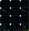
Immunolocalization of MPO, NO2Tyr, and fibronectin in rat aortic rings. Rat aortic rings were exposed to MPO (65 nM) for 120 minutes, washed, and incubated with NO2 (100 μM) and H2O2 (50 μM) for 90 minutes. In some cases, vessel explants were preincubated with enoxaparin (150 μg·ml–1) and washed prior to MPO exposure and omitting the washing step before H2O2 and NO2 addition. Tissue antigen distribution was visualized using rabbit polyclonal anti-MPO (green), mouse monoclonal anti-NO2Tyr (red), and rabbit polyclonal anti-rat fibronectin (green). (ac) Controls. Untreated vessel segments immunostained for MPO and NO2Tyr (a), vessel segments incubated with MPO and stained for NO2Tyr (b), and vessel segments treated with H2O2 and NO2 stained for NO2Tyr (c). (df) MPO and NO2Tyr distribution. Immunoreactivity for MPO (d) and NO2Tyr (e) colocalize, as shown when images were overlaid (f). (gi) Effect of enoxaparin on NO2Tyr formation. Untreated vessel stained for NO2Tyr (g), MPO-catalyzed NO2Tyr formation (h) was reduced when vessel explants were preincubated with enoxaparin (i). (jl) Vascular FN and NO2Tyr distribution. Subendothelial fibronectin immunoreactivity (j) colocalizes with NO2Tyr immunoreactivity (k), as shown in the merged image (l). L, vessel lumen; M, media. Arrows indicate endothelial cells. ×100 (af, jl); ×50 (gi).
Figure 7
Figure 7
Nitrotyrosine/tyrosine ratio in liver of MPO–/– mice after zymosan treatment. MPO–/– and wild-type mice (MPO+/+) received either intraperitoneal zymosan (n = 6), or sodium chloride (NaCl) as a control (n = 6). After 96 hours, the liver was perfused and processed for quantitative nitrotyrosine and tyrosine analysis as described in Methods. P < 0.05, MPO–/– zymosan-treated versus MPO+/+ injected with zymosan. *P < 0.05, MPO+/+ zymosan-treated versus untreated MPO+/+ control.

Similar articles

Cited by

References

    1. Ischiropoulos H. Biological tyrosine nitration: a pathophysiological function of nitric oxide and reactive oxygen species. Arch Biochem Biophys. 1998;356:1–11. - PubMed
    1. Eiserich JP, et al. Microtubule dysfunction by posttranslational nitrotyrosination of α-tubulin: a nitric oxide-dependent mechanism of cellular injury. Proc Natl Acad Sci USA. 1999;96:6365–6370. - PMC - PubMed
    1. Cassina AM, et al. Cytochrome c nitration by peroxynitrite. J Biol Chem. 2000;275:21409–21415. - PubMed
    1. Viner RI, Ferrington DA, Williams TD, Bigelow DJ, Schoneich C. Protein modification during biological aging: selective tyrosine nitration of the SERCA2a isoform of the sarcoplasmic reticulum Ca2+-ATPase in skeletal muscle. Biochem J. 1999;340:657–669. - PMC - PubMed
    1. MacMillan-Crow LA, Crow JP, Kerby JD, Beckman JS, Thompson JA. Nitration and inactivation of manganese superoxide dismutase in chronic rejection of human renal allografts. Proc Natl Acad Sci USA. 1996;93:11853–11858. - PMC - PubMed

Publication types