. 2001 Dec;108(12):1797-806.
doi: 10.1172/JCI13877.

Proteasomal regulation of betac signaling reveals a novel mechanism for cytokine receptor heterotypic desensitization

Affiliations

Proteasomal regulation of betac signaling reveals a novel mechanism for cytokine receptor heterotypic desensitization

M Martinez-Moczygemba et al. J Clin Invest. 2001 Dec.

Abstract

IL-5, IL-3, and GM-CSF are hematopoietic cytokines that are key mediators of the allergic inflammatory response. The receptors for these three cytokines consist of a cytokine-specific alpha (Ralpha) chain and a shared common beta (betac) chain. Herein, we demonstrate that agonistic ligation of these receptor subunits rapidly induces proteasomal degradation of the betac, but not the Ralpha, cytoplasmic domain, resulting in termination of signal transduction and yielding a truncated betac isoform ligated to the Ralpha subunit. Proteasomal degradation of the betac cytoplasmic domain was also a prerequisite for endocytosis and lysosomal degradation of the ligated receptor subunits. Moreover, proteasome-dependent termination of signaling induced by one betac-engaging cytokine resulted in cellular desensitization to signal transduction by subsequent stimulation with another betac-engaging cytokine. These data provide the first evidence for ligand-dependent proteasomal degradation of the betac cytoplasmic domain, and they establish a novel mechanism for heterotypic desensitization of shared cytokine receptor signaling.


Figures

Figure 1
Figure 1
IL-5 reduces βc protein expression, while transiently increasing faster-migrating βc bands. (a) TF1 cells that were cytokine-starved for 48 hours were stimulated with 5 ng/ml of either IL-5, GM-CSF, or IL-3 for the indicated times (107 cells/lane). Whole-cell lysates were prepared and immunoprecipitated with anti-βc monoclonal antibody S-16. Immune complexes were resolved on 8% SDS-PAGE gels and immunoblotted with anti-βc polyclonal antibodies. The top arrow in each panel represents full-length βc receptors, and the bottom arrow indicates truncated βc receptors (Δβc). (b) TF1 cells that were cytokine-starved for 48 hours were stimulated with 5 ng/ml of either IL-5, IL-3, or GM-CSF for the indicated times (107 cells/lane). Total RNA was prepared for each timepoint, and 30 μg of RNA/lane was analyzed with a βc probe corresponding to residues 417–530, which includes a piece of the membrane-proximal extracellular domain, the transmembrane domain, and part of the intracytoplasmic domain. (c) Total RNA from the IL-5–stimulated (for 1 hour) TF1 cells was reverse-transcribed using oligo(dT) primers, and amplified by PCR using a 3′ βc primer corresponding to its poly-A+ tail, a 5′ primer to a region flanking the βc transmembrane domain (lane 3), and another 5′ primer located in its transmembrane domain (lane 4). As a negative control, no cDNA was included in lane 2. Lane 1 is a DNA size ladder.
Figure 2
Figure 2
IL-5 induces proteasomal degradation of the βc cytoplasmic domain. (a) TF1 cells that were cytokine-starved for 24 hours were left untreated (lanes 1, 3, and 5) or were pretreated with 50 μM LLnL (lanes 2, 4, and 6) or 0.01% (vol) DMSO (lane 7) for 1 hour prior to IL-5 stimulation (10 ng/ml) (top and bottom panels). Whole-cell lysates were immunoprecipitated with anti-βc monoclonal antibody S-16, and immunoblotted with anti-βc polyclonal antibodies (top panel). The top arrow indicates full-length βc receptors; the bottom arrow corresponds to βIP. The membrane was stripped and reprobed with monoclonal antibody 4G10 (bottom panel). (b) Same as in a except the cells were pretreated with 20 μM β-lactone for 1 hour, followed by IL-5 stimulation. The top panel shows analysis by IP/IB with the same anti-βc antibodies described in a. The membrane was then stripped and reprobed with 4G10 (bottom panel). (c) Same as in a and b, except that the cells were pretreated with 50 μM MG132 for 1 hour, followed by IL-5 stimulation (all panels). The top panel was analyzed by IP/IB with the same anti-βc antibodies used in a and b. The middle panel was immunoprecipitated with 4G10, and immunoblotted with the anti-βc polyclonal antibodies. The blot was then stripped and reprobed with both anti-JAK2 and anti-STAT5 antibodies (bottom panel). (d) TF1 cells (3 × 10⁷ cells/IP) that had been cytokine-starved for 24 hours were pretreated with 30 μM β-lactone for 1 hour, followed by IL-5 stimulation (15 ng/ml). Whole-cell lysates were immunoprecipitated with anti-βc antibodies and immunoblotted with anti-ubiquitin monoclonal antibodies (left panel). The bracket designates βc ubiquitination, as well as other ubiquitinated proteins that coimmunoprecipitated with anti-βc antibodies. The blot in the left panel was stripped and reprobed with anti-βc antibodies (right panel). DM, DMSO, Yp, tyrosine phosphorylation; Ub, ubiquitin.
Figure 3
Figure 3
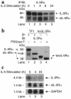
IL-5 regulation of transmembrane IL-5Rα and sIL-5Rα expression. (a) TF1 cells (107/lane) were cytokine-starved for 48 hours and stimulated with 5 ng/ml human IL-5 for the indicated times. Whole-cell lysates were prepared and subjected to IP/IB with anti–IL-5Rα antibodies. The top arrow represents IL-5Rα (60 kDa), and the bottom arrow corresponds to sIL-5Rα (50 kDa). The multiple bands represent differential glycosylation (see below). (b) TF1 cells continuously cultured in IL-5 were immunoprecipitated with anti–IL-5Rα antibodies (lanes 1 and 2), and either left untreated (lane 1) or treated with PNGase F (lane 2) for 1–2 hours to remove N-linked glycosyl groups. Lanes 3 and 4 are controls for sIL-5Rα migration, using 20 ng of baculovirus-expressed, recombinant sIL-5Rα protein (brsIL-5Rα), which was either untreated (lane 3) or treated with PNGase F (lane 4) for 1–2 hours, separated by 12.5% SDS-PAGE, and immunoblotted with anti–IL-5Rα antibodies. Note how the multiple bands in lane 1 resolve to two distinct bands in lane 2 (PNGase F–treated). (c) TF1 cells (107/lane) were cytokine-starved for 48 hours and stimulated with 5 ng/ml IL-5 for the indicated times. Total RNA was prepared for each timepoint, and 30 μg of RNA/lane were analyzed with a probe specific to the extracellular domain of IL-5Rα (detects both isoforms). GAPDH was used as an internal control.
Figure 4
Figure 4
Proteasome inhibitors stabilize IL-5Rα and βc protein/protein interaction. TF1 cells that were cytokine-starved for 48 hours were either left untreated (lanes 1, 3, and 5) or were pretreated with 50 μM LLnL (lanes 2, 4, and 6) or 0.01% (vol) DMSO (lane 7) for 1 hour prior to IL-5 stimulation (10 ng/ml) for the indicated times. Whole-cell lysates were prepared, and IP/IB analysis was performed with anti–IL-5Rα polyclonal antibody (top panel). The blot in the top panel was stripped and reprobed with anti-βc polyclonal antibodies (bottom panel). Note that both full-length βc receptor and βIP coimmunoprecipitate with IL-5Rα antibodies (bottom panel).
Figure 5
Figure 5
The ligated IL-5R complex is endocytosed and degraded in lysosomes. (a) TF1 cells that had been cytokine-starved for 24 hours were left untreated (lanes 1, 3, and 5) or were pretreated with 10 μM cytochalasin D (lanes 2, 4, and 6) for 1 hour prior to IL-5 stimulation (10 ng/ml) for the indicated times. Whole-cell lysates were prepared, and proteins were immunoprecipitated with anti-βc monoclonal antibody and immunoblotted with anti-βc polyclonal antibodies (top panel). The membrane was stripped and reprobed with anti–IL-5Rα antibodies (bottom panel). Note the accumulation of βc, βIP, and IL-5Rα in the presence of the inhibitor in lane 6. (b) Same as in a, top panel, except that cells were pretreated with 200 nM bafilomycin A1 in lanes 2, 4, and 6. Note the accumulation of βIP in the presence of bafilomycin A1 in lane 6 (bottom arrow).
Figure 6
Figure 6
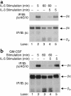
IL-5 stimulation results in cellular refractoriness to other βc-engaging cytokines. (a) TF1 cells that had been cytokine-starved for 24 hours were unstimulated (lane 1), or were stimulated with 20 ng/ml IL-5 for 5 minutes (lane 2) or 1 hour (lane 3). After 1 hour of incubation in IL-5, cells in lane 4 were harvested, washed once in 1× PBS (to remove IL-5), and restimulated with 20 ng/ml IL-3 for 5 minutes. As a positive control (lane 5), cells were stimulated only with 20 ng/ml IL-3 for 5 minutes. Whole-cell lysates were prepared and immunoprecipitated with anti-βc monoclonal antibody and immunoblotted with monoclonal antibody 4G10 (upper panel). The membrane was stripped and reprobed with anti-βc polyclonal antibodies (bottom panel). The migration of βc and βIP are indicated. (b) Same as in a, except that cells were pretreated with 20 ng/ml GM-CSF for 5 minutes or 1 hour, washed once in 1× PBS (to remove GM-CSF), and restimulated with 20 ng/ml IL-5 for 5 minutes. As a positive control (lane 5), cells were stimulated with 20 ng/ml IL-5 for 5 minutes only.
Figure 7
Figure 7
Model of IL-5R subunit regulation following IL-5 stimulation. IL-5 engagement of IL-5Rα leads to the recruitment of βc to form the high-affinity IL-5R (step 1). This protein/protein interaction (IL-5Rα/βc/IL-5) leads to the activation of intracellular signaling pathways that result in the activation of βc by both tyrosine and serine phosphorylation (step 2). Once activated, the cytoplasmic domains of βc are rapidly degraded by proteasomes to terminate agonistic signaling, resulting in the generation of βIP/IL-5Rα/IL-5 complexes (step 3). This molecular complex is then endocytosed (step 4) and degraded in lysosomes (steps 4 and 5). In addition, IL-5 induces a reduction in IL-5Rα mRNA and protein expression, while simultaneously increasing sIL-5Rα expression (upper left corner). S-p, serine phosphorylation.

Similar articles

Cited by

References

    1. Metcalf D. Hematopoietic regulators: redundancy or subtlety? Blood. 1993;82:3515–3523. - PubMed
    1. Arai KI, et al. Cytokines: coordinators of immune and inflammatory responses. Annu Rev Biochem. 1990;59:783–836. - PubMed
    1. Tavernier J, et al. A human high affinity interleukin-5 receptor (IL-5R) is composed of an IL-5-specific α chain and a β chain shared with the receptor for GM-CSF. Cell. 1991;66:1175–1184. - PubMed
    1. Kitamura T, Sato N, Arai K, Miyajima A. Expression cloning of the human IL-3 receptor cDNA reveals a shared β subunit for the human IL-3 and GM-CSF receptors. Cell. 1991;66:1165–1174. - PubMed
    1. Hayashida K, et al. Molecular cloning of a second subunit of the receptor for human granulocyte-macrophage colony-stimulating factor (GM-CSF): reconstitution of a high-affinity GM-CSF receptor. Proc Natl Acad Sci USA. 1990;87:9655–9659. - PMC - PubMed

Publication types

MeSH terms