. 2001 Dec;108(12):1807-16.
doi: 10.1172/JCI12218.

Cellular bicarbonate protects rat duodenal mucosa from acid-induced injury

Affiliations

Cellular bicarbonate protects rat duodenal mucosa from acid-induced injury

Y Akiba et al. J Clin Invest. 2001 Dec.

Abstract

Secretion of bicarbonate from epithelial cells is considered to be the primary mechanism by which the duodenal mucosa is protected from acid-related injury. Against this view is the finding that patients with cystic fibrosis, who have impaired duodenal bicarbonate secretion, are paradoxically protected from developing duodenal ulcers. Therefore, we hypothesized that epithelial cell intracellular pH regulation, rather than secreted extracellular bicarbonate, was the principal means by which duodenal epithelial cells are protected from acidification and injury. Using a novel in vivo microscopic method, we have measured bicarbonate secretion and epithelial cell intracellular pH (pH(i)), and we have followed cell injury in the presence of the anion transport inhibitor DIDS and the Cl(-) channel inhibitor, 5-nitro-2-(3-phenylpropylamino) benzoic acid (NPPB). DIDS and NPPB abolished the increase of duodenal bicarbonate secretion following luminal acid perfusion. DIDS decreased basal pH(i), whereas NPPB increased pH(i); DIDS further decreased pH(i) during acid challenge and abolished the pH(i) overshoot over baseline observed after acid challenge, whereas NPPB attenuated the fall of pH(i) and exaggerated the overshoot. Finally, acid-induced epithelial injury was enhanced by DIDS and decreased by NPPB. The results support the role of intracellular bicarbonate in the protection of duodenal epithelial cells from luminal gastric acid.


Figures

Figure 1
Figure 1
Effect of DIDS and NPPB on acid-induced bicarbonate secretion. Titratable alkalinity (bicarbonate secretion) was measured by the pH-stat method in duodenal loop perfusion experiments. Both DIDS and NPPB inhibit the increased bicarbonate secretion following acid exposure. All data are expressed as mean ± SEM from six rats. *P < 0.05 vs. pH 7.0 saline perfusion, P < 0.05 vs. pH 2.2 saline perfusion.
Figure 2
Figure 2
Effects of DIDS and NPPB on baseline pHi and on pHi during acid challenge of duodenal epithelial cells. (a) During perfusion with pH 7.0 solution, DIDS acidifies, whereas NPPB alkalinizes cells. (b) DIDS enhances, whereas NPPB attenuates acid-induced intracellular acidification during acid exposure. Note that DIDS abolishes, but NPPB enhances pHi overshoot following acid challenge. *P < 0.05 vs. pH 7.0 Krebs group, P < 0.05 vs. pH 2.2 Krebs group. All data are expressed as mean ± SEM from six rats.
Figure 3
Figure 3
Western blot of rat duodenum membrane proteins probed with pNBC1-specific (a) and kNBC1-specific (b) Ab’s. Rat duodenum membrane proteins (80 μg) were loaded in lanes 1 and 2 in both a and b. (a) Rat pancreas membrane proteins (25 μg)were loaded in lanes 3 and 4. (b) Rat kidney membrane proteins (100 μg) were loaded in lanes 3 and 4. The pNBC1-specific Ab and the kNBC1-specific Ab were used at dilution 1:1,000. For lanes 2 and 4, pNBC1 (a) and kNBC1 (b) Ab’s preincubated with the corresponding specific peptide (1:100) were used.
Figure 4
Figure 4
Immunolocalization of pNBC1 in rat duodenum. (a) pNBC1 is localized to the basolateral membranes of duodenal villus epithelial cells appearing as a cup-shaped structure (arrows). (b) When the pNBC1 Ab was preincubated with a specific peptide (1:100), staining was suppressed. (c) An image taken with Nomarski optics is included to provide orientation. The white rectangle encloses the same area shown in a. Calibration bar, 50 μm.
Figure 5
Figure 5
PI-positive staining in vivo and effect of acid. (a and d) A low number of visible PI-positive nuclei were visible 30 minutes after perfusion with pH 7.0 solution (t = 30 minutes). The red color of the villi is BCECF fluorescence observed through a 590-nm filter (thick arrows). (b and e) Fifteen minutes after a 5-minute pulse of pH 1.8 perfusate (t = 30 minutes), PI-positive dots are seen readily, localized to the villous tips (thin arrows). (c and f) Five minutes after a 5-minute pulse of pH 1.0 perfusate (t = 20 minutes), the villous tips have ballooned, and some are detached (yellow arrows). Note that all of the epithelial cells in the ballooned and detached villi are PI positive. Calibration bar, 100 μm. a, d, e, and f were printed so as to emphasize the faint staining of the villi.
Figure 6
Figure 6
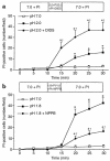
Effect of DIDS and NPPB on acid-induced duodenal injury (PI study). (a) Effect of DIDS. A 5-minute pulse of pH 2.0 injures the mucosa, as measured by a progressive increase of PI-positive nuclei. DIDS further increased the number of PI-positive cells induced by a pH 2.0 pulse. *P < 0.05 vs. pH 7.0 Krebs group, P < 0.05 vs. pH 2.0 Krebs group. (b) Effect of NPPB. NPPB lessens mucosal injury caused by a 5-minute perfusion with pH 1.8 solution, as assessed by decreased number of PI-positive cells. *P < 0.05 vs. pH 7.0 Krebs group, P < 0.05 vs. pH 1.8 Krebs group. All data are expressed as mean ± SEM from six rats.
Figure 7
Figure 7
Effect of DIDS and NPPB on acid-induced duodenal injury (H&E study). Injury was assessed in control and acid-exposed duodena with the use of conventional histologic sections. (a) Duodenal villi perfused with pH 7.0 solution, (b) pH 2.2 solution, (c) pH 2.0 solution, (d) pH 1.8 solution, (e) pH 2.0 + DIDS, and (f) pH 1.8 + NPPB. Note that DIDS increased mucosal injury, as manifest by focal necrosis seen in e (thin arrows), compared with no injury seen with pH 2.0 alone (c). The sloughing caused by pH 1.8 perfusion (thick arrows), seen in d, was not observed in the presence of NPPB (f), where ballooning (arrowheads), a more mild form of injury, was observed (H&E, ×400; calibration bar, 100 μm).

Comment in

References

    1. Rhodes J, Apsimon HT, Lawrie JH. pH of the contents of the duodenal bulb in relation to duodenal ulcer. Gut. 1966;7:502–508. - PMC - PubMed
    1. Paimela H, Kiviluoto T, Mustonen H, Kivilaakso E. Intracellular pH of isolated Necturus duodenal mucosa exposed to luminal acid. Gastroenterology. 1992;102:862–867. - PubMed
    1. Garner A, Flemström G, Allen A, Heylings JR, McQueen S. Gastric mucosal protective mechanisms: roles of epithelial bicarbonate and mucus secretions. Scand J Gastroenterol Suppl. 1984;101:79–86. - PubMed
    1. Isenberg JI, Selling JA, Hogan DL, Koss MA. Impaired proximal duodenal mucosal bicarbonate secretion in patients with duodenal ulcer. N Engl J Med. 1987;316:374–379. - PubMed
    1. Hogan DL, et al. Duodenal bicarbonate secretion: eradication of Helicobacter pylori and duodenal structure and function in humans. Gastroenterology. 1996;110:705–716. - PubMed

Publication types

MeSH terms

Substances