. 2001 Dec;108(12):1833-41.
doi: 10.1172/JCI13116.

Antisense inhibition of macrophage inflammatory protein 1-alpha blocks bone destruction in a model of myeloma bone disease

Affiliations

Antisense inhibition of macrophage inflammatory protein 1-alpha blocks bone destruction in a model of myeloma bone disease

S J Choi et al. J Clin Invest. 2001 Dec.

Abstract

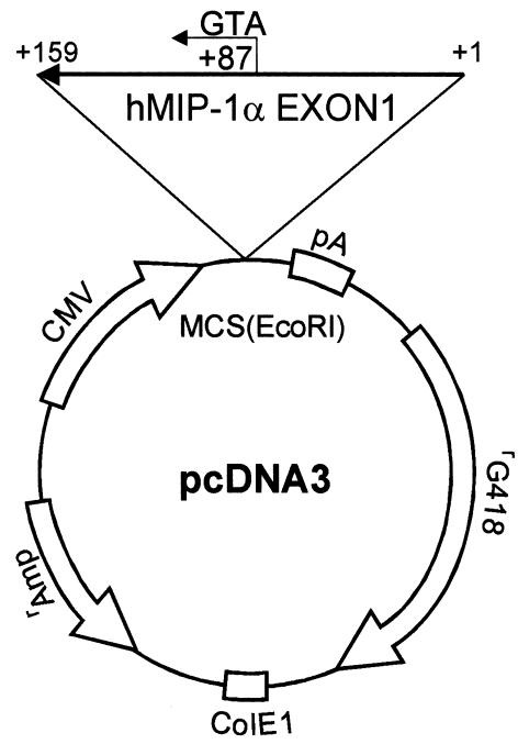
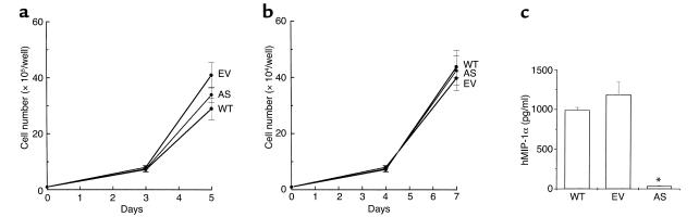
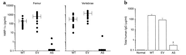
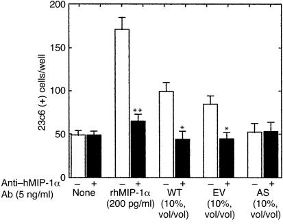
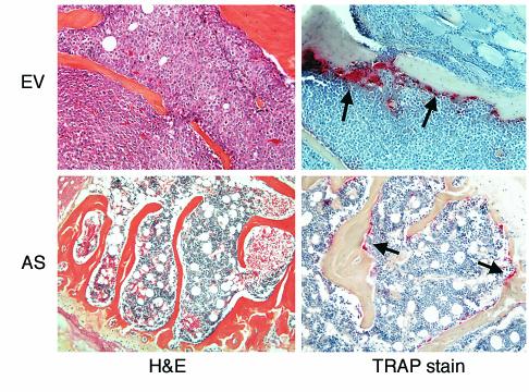
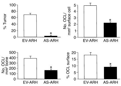
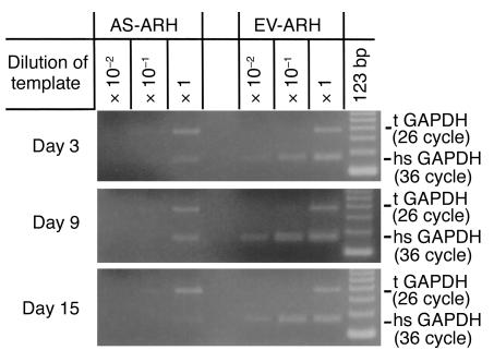
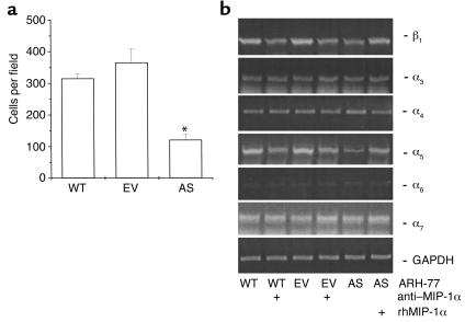
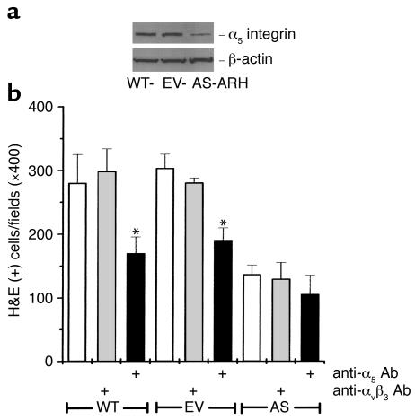
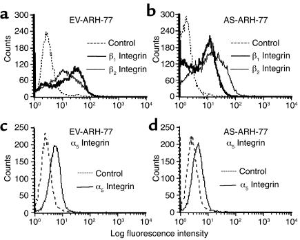
We recently identified macrophage inflammatory protein 1-alpha (MIP-1alpha) as a factor produced by multiple myeloma (MM) cells that may be responsible for the bone destruction in MM (1). To investigate the role of MIP-1alpha in MM bone disease in vivo, the human MM-derived cell line ARH was stably transfected with an antisense construct to MIP-1alpha (AS-ARH) and tested for its capacity to induce MM bone disease in SCID mice. Human MIP-1alpha levels in marrow plasma from AS-ARH mice were markedly decreased compared with controls treated with ARH cells transfected with empty vector (EV-ARH). Mice treated with AS-ARH cells lived longer than controls and, unlike the controls, they showed no radiologically identifiable lytic lesions. Histomorphometric analysis demonstrated that osteoclasts (OCLs) per square millimeter of bone and OCLs per millimeter of bone surface of AS-ARH mice were significantly less than in EV-ARH mice, and the percentage of tumors per total bone area was also significantly decreased. AS-ARH cells demonstrated decreased adherence to marrow stromal cells, due to reduced expression of the alpha(5)beta(1) integrin and diminished homing capacity and survival. These data support an important role for MIP-1alpha in cell homing, survival, and bone destruction in MM.


Figures

Figure 1
Figure 1
Construction of MIP-1α antisense. The first exon of MIP-1α cDNA was generated by standard PCR techniques as described in Methods. The identity of the MIP-1α cDNA was confirmed by sequence analysis, and it was subcloned into the pcDNA3 vector. Clones that had the reverse orientation for the MIP-1α cDNA were screened by PCR, and the orientation was confirmed by DNA sequence analysis.
Figure 2
Figure 2
Growth characteristics of WT-, EV-, and AS-ARH cells in vitro. (a) WT-, EV-, and AS-ARH cells (105) were cultured in six-well plates containing RPMI-1640 media containing 10% FBS. At days 3 and 5 of the culture, the cells were sampled, stained with trypan blue, and counted. (b) ST2 cells (104) in αMEM containing 10% FBS were plated onto dentin slices in 48-well plates. After 24 hours, WT-, EV-, and AS-ARH cells (104) in RPMI-1640 media containing 10% FBS were added to the culture. At days 4 and 7 of the culture, viable cells were scored as described above. Growth rates of WT-, EV-, and AS-ARH cells were not significantly different in the presence or absence of ST2 cells cocultured on dentin slices. (c) Conditioned media from WT-, EV-, or AS-ARH cells (105/ml) cultured in RPMI-1640 media containing 10% FBS were harvested at day 3, and the expression levels of MIP-1α were measured with a MIP-1α ELISA kit according to the manufacturer’s protocol. Similar results were seen in three independent experiments (*P < 0.0001).
Figure 3
Figure 3
Survival of SCID mice implanted with WT-, ET-, or AS-ARH cells and expression of MIP-1α in vivo. WT-, EV-, and AS-ARH cells were infused intravenously into SCID mice (n = 10 per group) as described in Methods and were sacrificed when they became paraplegic. Femurs and vertebrae were then removed and bone marrow plasma obtained by flushing the bones with 1 ml of serum-free αMEM. Expression levels of hMIP-1α (a) and human IgG (b) were measured with ELISA kits. hMIP-1α expression in mice implanted with AS-ARH cells was reduced to almost undetectable levels. Human IgG levels, which are indicators of tumor burden, were significantly reduced in AS-ARH mice compared with WT- or EV-ARH mice, but were still detectable (0.1–1 μg/ml). Similar results were seen in three independent experiments (*P < 0.0001).
Figure 4
Figure 4
Effect of bone marrow plasma from SCID mice implanted with WT-, ET-, or AS-ARH cells on human OCL-like formation. Bone marrow plasma (10% vol/vol) from SCID mice infused with WT-, ET-, or AS-ARH cells that were obtained at the time they developed paraplegia, was added to human bone marrow cultures in the absence of exogenously added osteoclastogenic factors. rhMIP-1α (200 pg/ml) stimulated OCL-like MNC formation, which was blocked by the MIP-1α–neutralizing Ab (5 ng/ml). Bone marrow plasma from WT- or EV-ARH mice stimulated OCL-like MNC formation that was blocked by the MIP-1α neutralizing Ab. In contrast, bone marrow plasma from AS-ARH mice did not stimulate OCL-like MNC formation. Similar results were seen in two independent experiments (*P < 0.05; **P < 0.01).
Figure 5
Figure 5
Histology of bone sections from SCID mice implanted with EV- or AS-ARH cells. As shown in the panels stained with hematoxylin and eosin (H&E), mice implanted with AS-ARH cells had significantly reduced tumor burden compared with mice implanted with EV-ARH cells. OCL number was markedly reduced in SCID mice implanted with AS-ARH cells compared with the mice implanted with EV-ARH cells.
Figure 6
Figure 6
Histomorphometric analysis of bone sections from SCID mice. Mice treated with AS-ARH cells had markedly reduced tumor burden compared with mice receiving EV-ARH cells. Furthermore, the number of OCLs per square millimeter of surface, the number of OCLs per square millimeter of bone area, and the percentage of active OCL surface were significantly reduced in AS-ARH mice compared with the EV-ARH mice (*P < 0.05).
Figure 7
Figure 7
Expression levels of human-specific GAPDH mRNA from SCID mice implanted with EV- or AS-ARH cells at 3 to 15 days after cell infusion. Mice were infused with EV- or AS-ARH as described in Methods and sacrificed at 3-day intervals. Marrow samples were harvested and tested for human-specific GAPDH (hsGAPDH) mRNA expression by serial dilution RT-PCR analysis. The amount of hsGAPDH present in the bone marrow of SCID mice at days 3, 9, and 15 was significantly decreased compared with that of EV-ARH. The hsGAPDH was detectable up to 15 days in bone marrow of SCID mice infused with AS-ARH cells. In contrast, hsGAPDH levels in mice transplanted with EV-ARH cells were at least two logs higher compared with that in mice infused with AS-ARH cells. GAPDH primers that could detect both murine and human GAPDH mRNA were used as an internal control (tGAPDH).
Figure 8
Figure 8
Adhesion of WT-, EV-, or AS-ARH cells to ST2 mouse marrow stromal cells and expression levels of human β1 integrin mRNAs. (a) WT-, EV-, or AS-ARH cells (106) in RPMI-1640 media containing 10% FBS were cocultured with ST2 mouse stromal cells (106) in six-well plates. After 4 days, the plates were extensively washed with 3 ml of serum-free RPMI-1640 media five times to remove nonadherent ARH cells. The cells were fixed with acetone followed by H&E staining. Blue-stained plasma cells attached onto ST2 cells were scored using an inverted microscope by counting ten random fields (×400). The number of AS-ARH cells attached onto ST2 mouse stromal cells was significantly reduced compared with the EV- or WT-ARH cells. (b) Expression levels of the mRNAs for the β1 integrin were measured as described in Methods. Expression levels for the human α3, α4, α6, α7 integrins mRNAs were not affected by MIP-1α. In contrast, mRNA expression levels for the human α5 and β1 integrins were significantly decreased in AS-ARH cells and increased two- to fourfold in AS-ARH cells treated with MIP-1α. Treatment of WT- or EV-ARH cells with an anti–MIP-1α Ab decreased mRNA expression of the human α5 and β1 integrins by approximately 50% (*P < 0.05). Similar results were seen in two independent experiments.
Figure 9
Figure 9
Expression of human integrin α5 in WT-, EV-, or AS-ARH cells and the effects of an anti-CD49e Ab (human integrin α5) on the adherence of ARH cells to ST2 mouse marrow stromal cells. (a) Western blot analysis for expression of the α5 integrin of VLA5 in WT-, EV-, and AS-ARH cells. Western blot analysis was performed as described in Methods. A greater than 70% reduction in α5 was seen in AS-ARH cells compared with WT- or EV-ARH cells. A similar pattern of results was seen in two independent experiments. (b) Effects of an Ab to the α5 integrin of VLA5 on adherence of ARH cells to ST2 marrow stromal cells. Adhesion assays were performed as described in Methods. Ab to α5 but not to αvβ3 blocked the adherence of WT- or EV-ARH cells to ST2 cells. A similar pattern of results was seen in two independent experiments. *P < 0.05.
Figure 10
Figure 10
Immunofluorescence staining for CD18, CD29, and CD49e. Cells were stained and analyzed as described in Methods. Staining for EV-ARH cells was compared with AS-ARH cells for a given Ab. For each Ab, cells were also stained with isotype-matched control Ab to correct for background staining. Twenty thousand cells were analyzed for each sample. (a and c) EV-ARH cells. (b and d) AS-ARH cells.

References

    1. Alsina M, et al. Development of an in vivo model of human multiple myeloma bone disease. Blood. 1996;87:1495–1501. - PubMed
    1. Choi SJ, et al. Macrophage inflammatory protein 1-alpha (MIP-1α) is a potential osteoclast stimulatory factor in multiple myeloma. Blood. 2000;96:671–675. - PubMed
    1. Han JH, Choi SJ, Kurihara N, Oba Y, Roodman GD. Macrophage inflammatory protein 1-alpha is an osteoclastogenic factor in myeloma that is independent of RANK ligand. Blood. 2001;97:3349–3353. - PubMed
    1. Kukita T, et al. Macrophage inflammatory protein-1α (LD78) expressed in human bone marrow: its role in regulation of hematopoiesis and osteoclast recruitment. Lab Invest. 1997;76:399–406. - PubMed
    1. Fuller K, Owens JM, Chambers TJ. Macrophage inflammatory protein-1-alpha and IL-18 stimulate the motility but suppress the resorption of isolated rat osteoclasts. J Immunol. 1995;154:6065–6072. - PubMed

Publication types