. 2001 Dec 17;194(12):1699-709.
doi: 10.1084/jem.194.12.1699.

Shear-dependent eosinophil transmigration on interleukin 4-stimulated endothelial cells: a role for endothelium-associated eotaxin-3

Affiliations

Shear-dependent eosinophil transmigration on interleukin 4-stimulated endothelial cells: a role for endothelium-associated eotaxin-3

S L Cuvelier et al. J Exp Med. .

Abstract

Leukocyte infiltration into inflammatory sites is regulated by the expression of adhesion and activation proteins, yet the role of these proteins in shear-dependent transmigration is poorly understood. We examined eosinophil recruitment on cytokine-stimulated human umbilical vein endothelial cells (HUVECs) under laminar flow conditions. Eosinophils rapidly transmigrated on interleukin (IL)-4-, but not TNF-stimulated HUVECs. Transmigration was shear dependent, with up to 90% of eosinophils transmigrating in the presence of shear and less than 25% of cells transmigrating under static conditions. Eosinophils express CC chemokine receptor CCR3 and are responsive to various CC chemokines. The effects of chemokines are mediated primarily through G(alpha)i, which is pertussis toxin sensitive. Greater than 65% of shear-dependent eosinophil transmigration on IL-4-stimulated HUVECs was blocked by either pertussis toxin or by an anti-CCR3 monoclonal antibody. Using reverse transcription polymerase chain reaction (RT-PCR) and Western blots, we found that IL-4-stimulated HUVECs produce both mRNA and protein for eotaxin-3. Eotaxin-3 was both released by HUVECs and expressed on the endothelial cell surface. Pretreatment of HUVECs with an anti-eotaxin-3 antibody blocked eosinophil transmigration to the same extent as an anti-CCR3 antibody. These results indicate that IL-4-stimulated HUVECs support shear-dependent eosinophil transmigration by upregulating eotaxin-3, and that surface association is critical for the role of eotaxin-3 in transmigration.


Figures

Figure 1.
Figure 1.
Eosinophils transmigrate across IL-4-stimulated HUVECs. Tightly confluent monolayers of HUVECs were stimulated for 24 h with M199/A alone (Control) or M199/A containing 20 ng/ml IL-4 (IL-4). Alternatively, cells were stimulated for 6 h with M199/A containing 20 ng/ml TNF (TNF). After the incubation, the flow chamber was assembled and isolated eosinophils (5 × 10⁵/ml) were perfused over the HUVECs for 4 min at a wall shear stress of 2 dyn/cm2. (A) Accumulation and (B) transmigration were quantified as described in Materials and Methods. Data are mean ± SEM of between 3 and 15 experiments. *P < 0.001. Representative images of interactions between eosinophils and (C) TNF-stimulated HUVECs (TNF) or eosinophils and (D) IL-4-stimulated HUVECs (IL-4) were digitally captured at 400× magnification. Eosinophils bound to TNF-stimulated HUVECs remained phase-bright, whereas eosinophils bound to IL-4–stimulated HUVECs changed shape and transmigrated, becoming phase-dark.
Figure 1.
Figure 1.
Eosinophils transmigrate across IL-4-stimulated HUVECs. Tightly confluent monolayers of HUVECs were stimulated for 24 h with M199/A alone (Control) or M199/A containing 20 ng/ml IL-4 (IL-4). Alternatively, cells were stimulated for 6 h with M199/A containing 20 ng/ml TNF (TNF). After the incubation, the flow chamber was assembled and isolated eosinophils (5 × 10⁵/ml) were perfused over the HUVECs for 4 min at a wall shear stress of 2 dyn/cm2. (A) Accumulation and (B) transmigration were quantified as described in Materials and Methods. Data are mean ± SEM of between 3 and 15 experiments. *P < 0.001. Representative images of interactions between eosinophils and (C) TNF-stimulated HUVECs (TNF) or eosinophils and (D) IL-4-stimulated HUVECs (IL-4) were digitally captured at 400× magnification. Eosinophils bound to TNF-stimulated HUVECs remained phase-bright, whereas eosinophils bound to IL-4–stimulated HUVECs changed shape and transmigrated, becoming phase-dark.
Figure 2.
Figure 2.
Eosinophil transmigration across IL-4–stimulated HUVECs is shear dependent. HUVECs were stimulated with M199/A containing 20 ng/ml IL-4 for 24 h. Eosinophils (5 × 10⁵/ml) were allowed to interact with IL-4–stimulated HUVECs under static (0 dyn/cm2) or flow (2 dyn/cm2) conditions in the flow chamber. (A) Transmigration was measured every 2 min for 20 min as described in Materials and Methods. Data are mean ± SEM of at least four experiments. *P < 0.05. (B) Digitally captured images are presented at 1-min intervals to illustrate the interactions between eosinophils (black arrow) and IL-4–stimulated HUVECs under static or shear conditions. Data are representative of at least four experiments.
Figure 2.
Figure 2.
Eosinophil transmigration across IL-4–stimulated HUVECs is shear dependent. HUVECs were stimulated with M199/A containing 20 ng/ml IL-4 for 24 h. Eosinophils (5 × 10⁵/ml) were allowed to interact with IL-4–stimulated HUVECs under static (0 dyn/cm2) or flow (2 dyn/cm2) conditions in the flow chamber. (A) Transmigration was measured every 2 min for 20 min as described in Materials and Methods. Data are mean ± SEM of at least four experiments. *P < 0.05. (B) Digitally captured images are presented at 1-min intervals to illustrate the interactions between eosinophils (black arrow) and IL-4–stimulated HUVECs under static or shear conditions. Data are representative of at least four experiments.
Figure 3.
Figure 3.
Role of ERK1/2 MAP kinase and endothelial cell permeability in shear-dependent eosinophil transmigration. HUVECs were stimulated with M199/A containing 20 ng/ml IL-4 for 24 h. (A) HUVECs were pretreated with 20 μM PD 98059 or an equivalent amount of DMSO for 30 min before the assembly of the flow chamber. Transmigration was assessed as described in Fig. 1. (B) The flow chamber was assembled and buffer was perfused across the surface at 2 dyn/cm2 for 5 min. Eosinophils were then drawn into the chamber, the flow was stopped, and transmigration was determined every 2 min for 20 min as described in Fig. 2. (C) IL-4–stimulated HUVECs were treated with 10 μM histamine for 5 min before the assembly of the flow chamber. Eosinophils were allowed to interact with IL-4–stimulated HUVECs under static (0 dyn/cm2) or flow (2 dyn/cm2) conditions in the flow chamber and transmigration was assessed as described in Fig. 2. Data are mean ± SEM of at least three experiments. *P < 0.05.
Figure 4.
Figure 4.
Eosinophil transmigration across IL-4–stimulated HUVECs is pertussis toxin sensitive and CCR3 dependent. HUVECs were stimulated with IL-4 as described in Fig. 1. Eosinophils were pretreated with 250 ng/ml pertussis toxin for 1 h (PTX). Alternatively, eosinophils were pretreated for 10 min with the following antibodies: 5 μg/ml anti-α4-integrin mAb, 5 μg/ml anti-β2-integrin mAb, both anti-α4-integrin mAb and anti-β2-integrin mAb, or 10 μg/ml anti-CCR3 mAb (CCR3). The flow chamber was assembled and eosinophil transmigration was assessed as described in Fig. 1. Data are mean ± SEM of at least three experiments. *P < 0.001; ns, not significant.
Figure 5.
Figure 5.
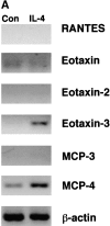
IL-4–stimulated HUVECs express eotaxin-3 mRNA and protein. HUVECs were stimulated for 24 h with M199/A alone or M199/A containing 20 ng/ml IL-4. (A) RNA was isolated from HUVECs using the TRIzol method and cDNA was generated using a First Strand synthesis kit. PCR was performed on cDNA using primers specific for CCR3 chemokines. Products were separated on agarose gels and visualized using ethidium bromide. β-actin served as a positive control. (B) HUVECs were lysed in a buffer containing Triton X-100 and lysates were separated by SDS-PAGE. Proteins were transferred to PVDF and eotaxin-3 protein was detected using an eotaxin-3-specific antibody. Figures are representative of at least three experiments.
Figure 5.
Figure 5.
IL-4–stimulated HUVECs express eotaxin-3 mRNA and protein. HUVECs were stimulated for 24 h with M199/A alone or M199/A containing 20 ng/ml IL-4. (A) RNA was isolated from HUVECs using the TRIzol method and cDNA was generated using a First Strand synthesis kit. PCR was performed on cDNA using primers specific for CCR3 chemokines. Products were separated on agarose gels and visualized using ethidium bromide. β-actin served as a positive control. (B) HUVECs were lysed in a buffer containing Triton X-100 and lysates were separated by SDS-PAGE. Proteins were transferred to PVDF and eotaxin-3 protein was detected using an eotaxin-3-specific antibody. Figures are representative of at least three experiments.
Figure 6.
Figure 6.
Eotaxin-3 protein is secreted by and associated with the surface of IL-4–stimulated HUVECs. HUVECs were stimulated for 24 h with M199/A alone or M199/A containing 20 ng/ml IL-4. (A) Supernatants were collected, and soluble eotaxin-3 was measured using a sandwich ELISA as described in Materials and Methods. (B) Surface-associated eotaxin-3 was detected using a cell-surface ELISA with nonimmune goat antibody (NI IgG) and anti-eotaxin-3 antibody as described in Materials and Methods. Data are mean ± SEM of at least five experiments. *P < 0.05; ns, not significant.
Figure 7.
Figure 7.
Surface-associated eotaxin-3 is responsible for the CCR3-dependent transmigration of eosinophils across IL-4–stimulated HUVECs. HUVECs were stimulated with IL-4 as described in Fig. 1. Freshly isolated eosinophils (5 × 10⁵/ml) were preincubated with 10 μg/ml of antibody against CCR3. In some experiments, HUVECs were preincubated for 10 min with 20 μg/ml of antibody against eotaxin-3 and then 10 μg/ml of eotaxin-3 was maintained in the perfusate. Isotype-matched control antibodies were used in all cases. The flow chamber was assembled and eosinophils were perfused over the HUVECs as described in Fig. 1. Transmigration was measured as described in Materials and Methods. Data are mean ± SEM of at least three experiments. *P < 0.001 compared with control and not significant compared with Eotaxin-3 or CCR3 alone.

Similar articles

Cited by

References

    1. Frigas, E., S. Motojima, and G.J. Gleich. 1991. The eosinophilic injury to the mucosa of the airways in the pathogenesis of bronchial asthma. Eur. Respir. J. (Suppl. 13):123s–135s. - PubMed
    1. Gleich, G.J. 1990. The eosinophil and bronchial asthma: current understanding. J. Allergy Clin. Immunol. 85:422–436. - PubMed
    1. Wardlaw, A.J., R. Moqbel, and A.B. Kay. 1995. Eosinophils: biology and role in disease. Adv. Immunol. 60:151–266. - PubMed
    1. Spertini, O., F.W. Luscinskas, M.A. Gimbrone, Jr., and T.F. Tedder. 1992. Monocyte attachment to activated human vascular endothelium in vitro is mediated by leukocyte adhesion molecule-1 (L-selectin) under nonstatic conditions. J. Exp. Med. 175:1789–1792. - PMC - PubMed
    1. Gopalan, P.K., A.R. Burns, S.I. Simon, S. Sparks, L.V. McIntire, and C.W. Smith. 2000. Preferential sites for stationary adhesion of neutrophils to cytokine-stimulated HUVEC under flow conditions. J. Leukoc. Biol. 68:47–57. - PubMed

Publication types