

Activation of natural killer T cells potentiates or prevents experimental autoimmune encephalomyelitis
- PMID: 11748280
- PMCID: PMC2193586
- DOI: 10.1084/jem.194.12.1789
Activation of natural killer T cells potentiates or prevents experimental autoimmune encephalomyelitis
Abstract
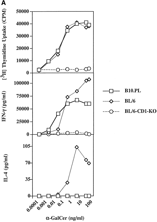
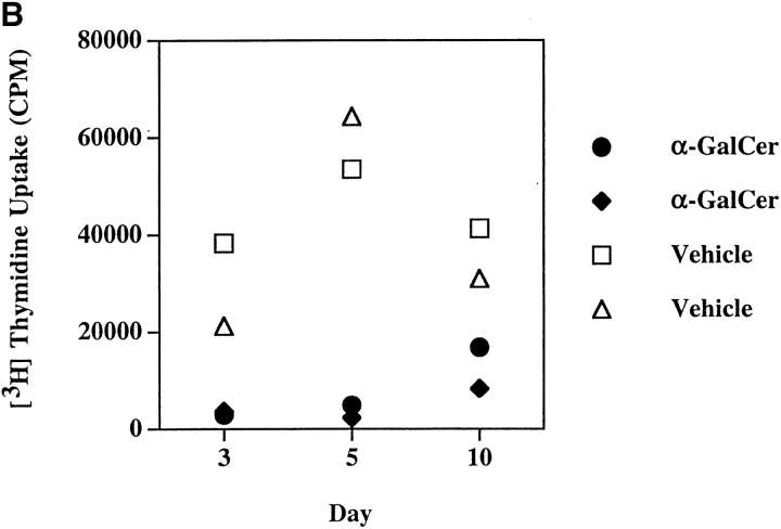
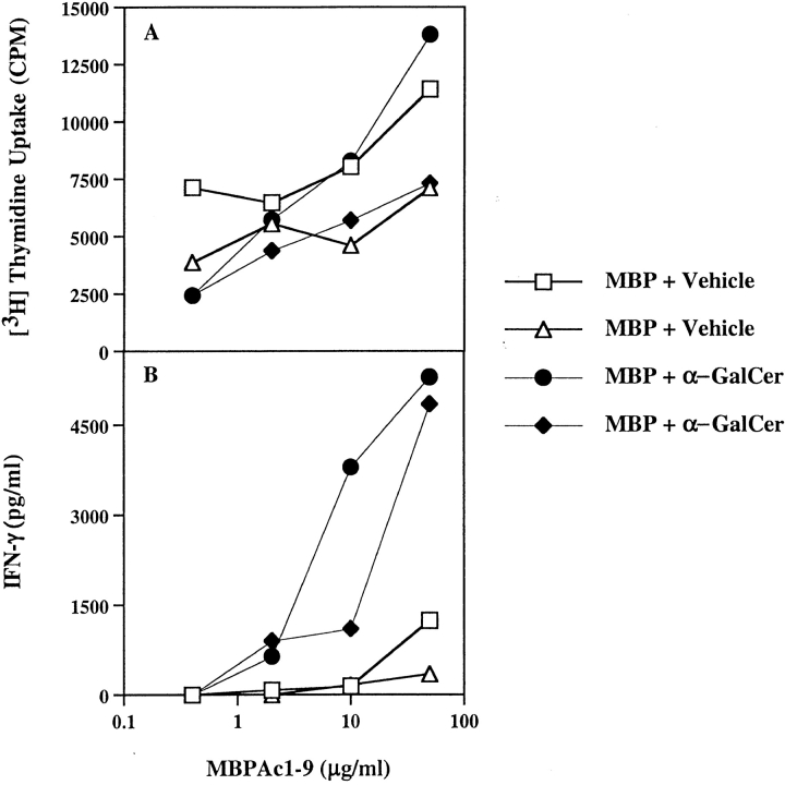
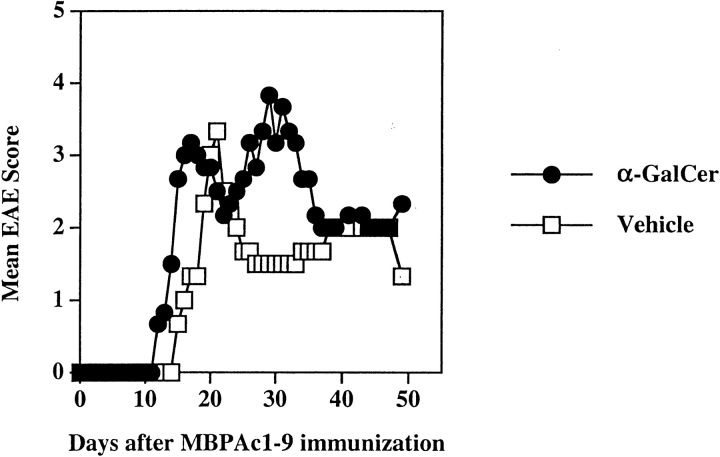
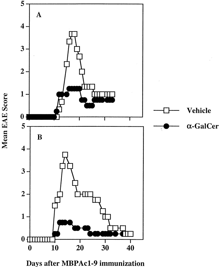
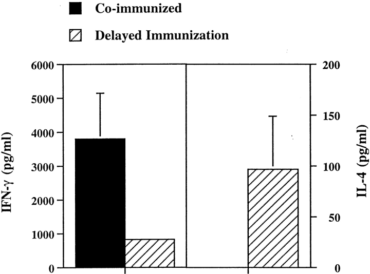
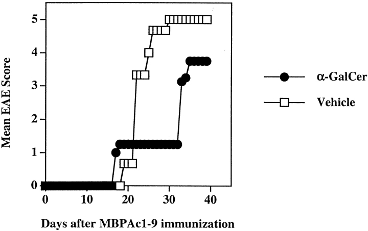
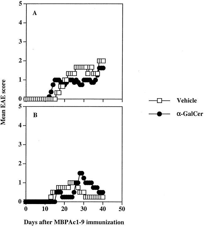
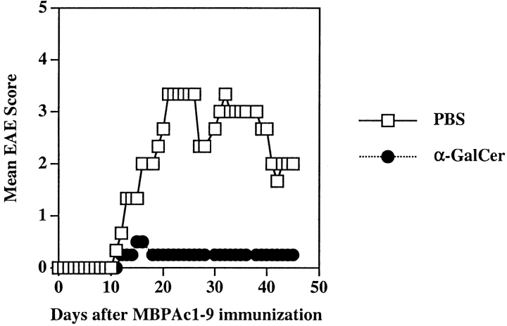
Natural killer (NK) T cells recognize lipid antigens in the context of the major histocompatibility complex (MHC) class 1-like molecule CD1 and rapidly secrete large amounts of the cytokines interferon (IFN)-gamma and interleukin (IL)-4 upon T cell receptor (TCR) engagement. We have asked whether NK T cell activation influences adaptive T cell responses to myelin antigens and their ability to cause experimental autoimmune encephalomyelitis (EAE), a model for multiple sclerosis. While simultaneous activation of NK T cells with the glycolipid alpha-galactosylceramide (alpha-GalCer) and myelin-reactive T cells potentiates EAE in B10.PL mice, prior activation of NK T cells protects against disease. Exacerbation of EAE is mediated by an enhanced T helper type 1 (Th1) response to myelin basic protein and is lost in mice deficient in IFN-gamma. Protection is mediated by immune deviation of the anti-myelin basic protein (MBP) response and is dependent upon the secretion of IL-4. The modulatory effect of alpha-GalCer requires the CD1d antigen presentation pathway and is dependent upon the nature of the NK T cell response in B10.PL or C57BL/6 mice. Because CD1 molecules are nonpolymorphic and remarkably conserved among different species, modulation of NK T cell activation represents a target for intervention in T cell-mediated autoimmune diseases.
Figures

Similar articles
-
Natural killer T cell activation protects mice against experimental autoimmune encephalomyelitis.J Exp Med. 2001 Dec 17;194(12):1801-11. doi: 10.1084/jem.194.12.1801. J Exp Med. 2001. PMID: 11748281 Free PMC article.
-
Abrogation of Endogenous Glycolipid Antigen Presentation on Myelin-Laden Macrophages by D-Sphingosine Ameliorates the Pathogenesis of Experimental Autoimmune Encephalomyelitis.Front Immunol. 2019 Mar 19;10:404. doi: 10.3389/fimmu.2019.00404. eCollection 2019. Front Immunol. 2019. PMID: 30941120 Free PMC article.
-
Costimulation-dependent modulation of experimental autoimmune encephalomyelitis by ligand stimulation of V alpha 14 NK T cells.J Immunol. 2001 Jan 1;166(1):662-8. doi: 10.4049/jimmunol.166.1.662. J Immunol. 2001. PMID: 11123351
-
Regulation of immune responses by CD1d-restricted natural killer T cells.Immunol Res. 2004;30(2):139-53. doi: 10.1385/IR:30:2:139. Immunol Res. 2004. PMID: 15477656 Review.
-
Lipid antigen presentation in the immune system: lessons learned from CD1d knockout mice.Immunol Rev. 1999 Jun;169:31-44. doi: 10.1111/j.1600-065x.1999.tb01304.x. Immunol Rev. 1999. PMID: 10450506 Review.
Cited by
-
Adjuvants of immunity: harnessing innate immunity to promote adaptive immunity.J Exp Med. 2002 Mar 4;195(5):F19-23. doi: 10.1084/jem.20020073. J Exp Med. 2002. PMID: 11877490 Free PMC article. No abstract available.
-
Lithium prevents and ameliorates experimental autoimmune encephalomyelitis.J Immunol. 2008 Jul 1;181(1):338-45. doi: 10.4049/jimmunol.181.1.338. J Immunol. 2008. PMID: 18566399 Free PMC article.
-
Epigenetic reduction in invariant NKT cells following in utero vitamin D deficiency in mice.J Immunol. 2011 Feb 1;186(3):1384-90. doi: 10.4049/jimmunol.1002545. Epub 2010 Dec 29. J Immunol. 2011. PMID: 21191070 Free PMC article.
-
A Novel Glycolipid Antigen for NKT Cells That Preferentially Induces IFN-γ Production.J Immunol. 2015 Aug 1;195(3):924-33. doi: 10.4049/jimmunol.1500070. Epub 2015 Jun 15. J Immunol. 2015. PMID: 26078271 Free PMC article.
-
Activated invariant NKT cells control central nervous system autoimmunity in a mechanism that involves myeloid-derived suppressor cells.J Immunol. 2013 Mar 1;190(5):1948-60. doi: 10.4049/jimmunol.1201718. Epub 2013 Jan 23. J Immunol. 2013. PMID: 23345328 Free PMC article.
References
-
- Bendelac, A., M.N. Rivera, S.H. Park, and J.H. Roark. 1997. Mouse CD1-specific NK1 T cells: development, specificity, and function. Annu. Rev. Immunol. 15:535–562. - PubMed
-
- Porcelli, S.A., and R.L. Modlin. 1999. The CD1 system: antigen-presenting molecules for T cell recognition of lipids and glycolipids. Annu. Rev. Immunol. 17:297–329. - PubMed
-
- Kawano, T., J. Cui, Y. Koezuka, I. Toura, Y. Kaneko, K. Motoki, H. Ueno, R. Nakagawa, H. Sato, E. Kondo, et al. 1997. CD1d-restricted and TCR-mediated activation of valpha14 NKT cells by glycosylceramides. Science. 278:1626–1629. - PubMed
Publication types
MeSH terms
Substances
LinkOut - more resources
Full Text Sources
Other Literature Sources
Research Materials
Miscellaneous
