. 2001 Dec 17;194(12):1813-21.
doi: 10.1084/jem.194.12.1813.

The extracellular domain of CD83 inhibits dendritic cell-mediated T cell stimulation and binds to a ligand on dendritic cells

Affiliations

The extracellular domain of CD83 inhibits dendritic cell-mediated T cell stimulation and binds to a ligand on dendritic cells

M Lechmann et al. J Exp Med. .

Abstract

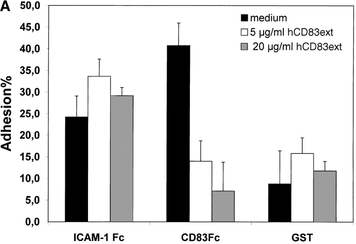
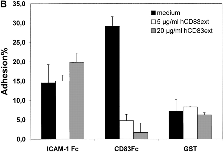
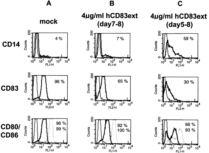
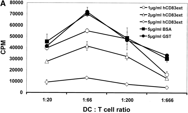
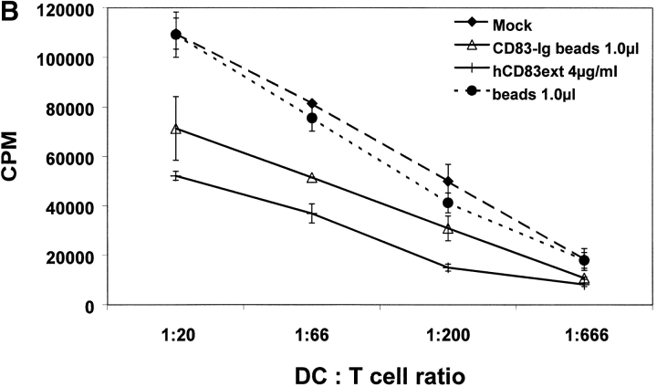
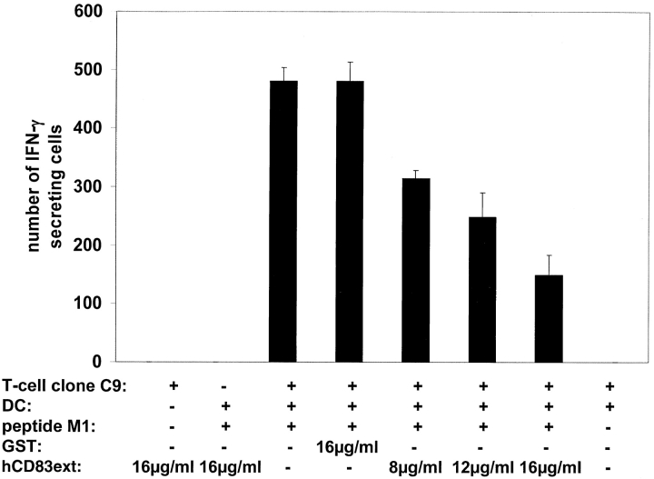
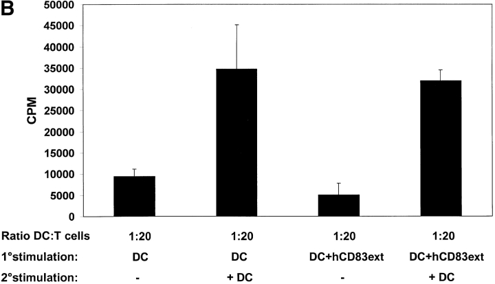
CD83 is an immunoglobulin (Ig) superfamily member that is upregulated during the maturation of dendritic cells (DCs). It has been widely used as a marker for mature DCs, but its function is still unknown. To approach its potential functional role, we have expressed the extracellular Ig domain of human CD83 (hCD83ext) as a soluble protein. Using this tool we could show that immature as well as mature DCs bind to CD83. Since CD83 binds a ligand also expressed on immature DCs, which do not express CD83, indicates that binding is not a homophilic interaction. In addition we demonstrate that hCD83ext interferes with DC maturation downmodulating the expression of CD80 and CD83, while no phenotypical effects were observed on T cells. Finally, we show that hCD83ext inhibits DC-dependent allogeneic and peptide-specific T cell proliferation in a concentration dependent manner in vitro. This is the first report regarding functional aspects of CD83 and the binding of CD83 to DCs.


Figures

Figure 1.
Figure 1.
Biophysical analyses of recombinant hCD83ext. (A) hCD83ext was separated by SDS-PAGE and silver stained. (B) Western blot analysis of the blotted hCD83ext using monoclonal anti-CD83 antibodies and (C) one-dimensional NMR spectrum, showing that hCD83ext is folded.
Figure 1.
Figure 1.
Biophysical analyses of recombinant hCD83ext. (A) hCD83ext was separated by SDS-PAGE and silver stained. (B) Western blot analysis of the blotted hCD83ext using monoclonal anti-CD83 antibodies and (C) one-dimensional NMR spectrum, showing that hCD83ext is folded.
Figure 1.
Figure 1.
Biophysical analyses of recombinant hCD83ext. (A) hCD83ext was separated by SDS-PAGE and silver stained. (B) Western blot analysis of the blotted hCD83ext using monoclonal anti-CD83 antibodies and (C) one-dimensional NMR spectrum, showing that hCD83ext is folded.
Figure 2.
Figure 2.
Plate adhesion assay. ICAM-Fc and hCD83-Fc were bound to goat anti–human Fc-specific F(ab′)2 precoated plates. Calcein-AM labeled DCs (40,000) were allowed to adhere for 45 min at 37°C. Fluorescence of adherent cells was quantified using fluorometric analyses. Clearly, immature (A) as well as mature (B) DCs bind to CD83. Preincubation of labeled DCs with recombinant hCD83ext inhibited the binding of the immature and mature DCs to plate-bound CD83-Fc. The binding could be inhibited in a concentration-dependent manner (5 and 20 μg/ml hCD83ext). Binding to ICAM-1-Fc as a control could not be inhibited by hCD83ext, while GST was used as a negative control. Both were not specifically influenced by the addition of hCD83ext. In addition the binding of DCs to CD83 was concentration dependent (C). The binding was lost when decreasing amounts of CD83-Fc were used. In these experiments, Ig coated via goat anti–human-Fc, was used as negative control. The Ig binding was not influenced by hCD83ext. These experiments were performed five times using different donors for DC generation. This represents a typical experiment.
Figure 2.
Figure 2.
Plate adhesion assay. ICAM-Fc and hCD83-Fc were bound to goat anti–human Fc-specific F(ab′)2 precoated plates. Calcein-AM labeled DCs (40,000) were allowed to adhere for 45 min at 37°C. Fluorescence of adherent cells was quantified using fluorometric analyses. Clearly, immature (A) as well as mature (B) DCs bind to CD83. Preincubation of labeled DCs with recombinant hCD83ext inhibited the binding of the immature and mature DCs to plate-bound CD83-Fc. The binding could be inhibited in a concentration-dependent manner (5 and 20 μg/ml hCD83ext). Binding to ICAM-1-Fc as a control could not be inhibited by hCD83ext, while GST was used as a negative control. Both were not specifically influenced by the addition of hCD83ext. In addition the binding of DCs to CD83 was concentration dependent (C). The binding was lost when decreasing amounts of CD83-Fc were used. In these experiments, Ig coated via goat anti–human-Fc, was used as negative control. The Ig binding was not influenced by hCD83ext. These experiments were performed five times using different donors for DC generation. This represents a typical experiment.
Figure 3.
Figure 3.
FACS® analyses of DCs. (A) Immature DCs were matured in the presence of the maturation cocktail from day 5–8 (= mock control for mature DCs). (B) Immature DCs where matured in the presence of the maturation cocktail (day 5–8) and on day 7 hCD83ext was added for 24 h. (C) Immature DCs where incubated in the presence of the maturation cocktail in combination with hCD83 from day 5–8. On day 8 cells where washed and stained with the indicated antibodies and analyzed by FACS®.
Figure 4.
Figure 4.
hCD83ext inhibits allogeneic T cell proliferation. (A) MLR analyses: the prokaryotic expressed hCD83ext reduced T cell proliferation in a dose-dependent manner. GST, which was purified in the same way as hCD83ext and BSA (each 5 μg/ml) were used as controls. (B) Also the eukaryotic expressed CD83–Fc fusion protein couplet to beads inhibits T cell proliferation. Uncoupled beads alone were used as a negative control and hCD83ext as a positive control. (C) The inhibition is not due to a contamination: to prove that the purified hCD83ext protein was responsible for the inhibitory activity and not a possible contamination present in the protein preparation, purified hCD83ext was filtered using a 10-kD Microcon system. Filtered and unfiltered hCD83ext exhibit comparable inhibitory effects. Whereas, cells which were treated with the filtrate alone showed no inhibition and the stimulation was comparable to mock-treated cells. All experiments were performed at least three times. Data presented here represent a typical experiment.
Figure 4.
Figure 4.
hCD83ext inhibits allogeneic T cell proliferation. (A) MLR analyses: the prokaryotic expressed hCD83ext reduced T cell proliferation in a dose-dependent manner. GST, which was purified in the same way as hCD83ext and BSA (each 5 μg/ml) were used as controls. (B) Also the eukaryotic expressed CD83–Fc fusion protein couplet to beads inhibits T cell proliferation. Uncoupled beads alone were used as a negative control and hCD83ext as a positive control. (C) The inhibition is not due to a contamination: to prove that the purified hCD83ext protein was responsible for the inhibitory activity and not a possible contamination present in the protein preparation, purified hCD83ext was filtered using a 10-kD Microcon system. Filtered and unfiltered hCD83ext exhibit comparable inhibitory effects. Whereas, cells which were treated with the filtrate alone showed no inhibition and the stimulation was comparable to mock-treated cells. All experiments were performed at least three times. Data presented here represent a typical experiment.
Figure 5.
Figure 5.
hCD83ext inhibits proliferation of influenza matrix peptide M1-specific CTL clone. DCs were loaded with influenza matrix peptide M1 and used to assess the stimulation (IFN-γ production) of the M1-specific CTL clone C9. Number of IFN-γ–producing spot are shown on the y-axis. Increasing concentrations of hCD83ext clearly reduced the number of spot forming cells. GST, which was purified in the same way as hCD83ext was used a negative control. Three independent experiment were performed. The data presented represent a typical experiment.
Figure 6.
Figure 6.
Proliferation arrest of hCD83ext-treated T cells can be restored. (A) hCD83ext-treated T cells derived from the primary MLRs, which have been either treated with or without hCD83ext, were stimulated for a second time period of 4 d without the addition of hCD83ext. Medium containing IL-2 or medium without IL-2 was added to the cultures. After a 3-d incubation period proliferation was determined. T cells which were strongly inhibited by hCD83ext in the primary MLRs could be restimulated with IL-2, clearly showing that the proliferation arrest, caused by hCD83ext in the primary MLRs, is not due to an irreversible toxic effect. (B) T cells which were inhibited with hCD83ext (4 μg/ml) in the primary MLRs (ratio DC:T cell = 1:20), were restimulated with allogeneic DCs (ratio 1:20). Arrested T cells could be restimulated with DCs, reaching stimulation levels comparable to those obtained with untreated cells in primary MLRs. Thus, the inhibitory function of hCD83ext is reversible. Experiments were performed at least three times. The data presented here represent a typical experiment.
Figure 6.
Figure 6.
Proliferation arrest of hCD83ext-treated T cells can be restored. (A) hCD83ext-treated T cells derived from the primary MLRs, which have been either treated with or without hCD83ext, were stimulated for a second time period of 4 d without the addition of hCD83ext. Medium containing IL-2 or medium without IL-2 was added to the cultures. After a 3-d incubation period proliferation was determined. T cells which were strongly inhibited by hCD83ext in the primary MLRs could be restimulated with IL-2, clearly showing that the proliferation arrest, caused by hCD83ext in the primary MLRs, is not due to an irreversible toxic effect. (B) T cells which were inhibited with hCD83ext (4 μg/ml) in the primary MLRs (ratio DC:T cell = 1:20), were restimulated with allogeneic DCs (ratio 1:20). Arrested T cells could be restimulated with DCs, reaching stimulation levels comparable to those obtained with untreated cells in primary MLRs. Thus, the inhibitory function of hCD83ext is reversible. Experiments were performed at least three times. The data presented here represent a typical experiment.

References

    1. Steinman, R.M. 1991. The dendritic cell system and its role in immunogenicity. Annu. Rev. Immunol. 9:271–296. - PubMed
    1. Banchereau, J., and R.M. Steinman. 1998. Dendritic cells and the control of immunity. Nature. 392:245–252. - PubMed
    1. Dieu, M.C., B. Vanbervliet, A. Vicari, J.M. Bridon, E. Oldham, S. Ait-Yahia, F. Briere, A. Zlotnik, S. Lebecque, and C. Caux. 1998. Selective recruitment of immature and mature dendritic cells by distinct chemokines expressed in different anatomic sites. J. Exp. Med. 188:373–386. - PMC - PubMed
    1. Zhou, L.J., R. Schwarting, H.M. Smith, and T.F. Tedder. 1992. A novel cell-surface molecule expressed by human interdigitating reticulum cells, Langerhans cells, and activated lymphocytes is a new member of the Ig superfamily. J. Immunol. 149:735–742. - PubMed
    1. Kozlow, E.J., G.L. Wilson, C.H. Fox, and J.H. Kehrl. 1993. Subtractive cDNA cloning of a novel member of the Ig gene superfamily expressed at high levels in activated B lymphocytes. Blood. 81:454–461. - PubMed

Publication types

MeSH terms