

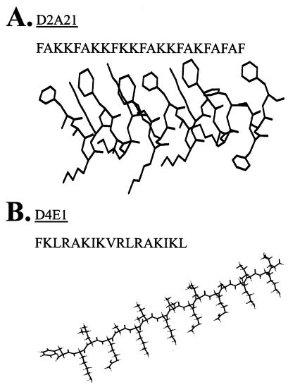
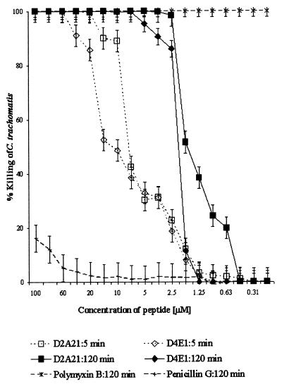
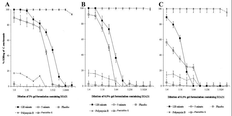
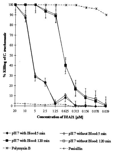
In vitro microbicidal activities of cecropin peptides D2A21 and D4E1 and gel formulations containing 0.1 to 2% D2A21 against Chlamydia trachomatis
- PMID: 11751108
- PMCID: PMC126975
- DOI: 10.1128/AAC.46.1.34-41.2002
In vitro microbicidal activities of cecropin peptides D2A21 and D4E1 and gel formulations containing 0.1 to 2% D2A21 against Chlamydia trachomatis
Abstract
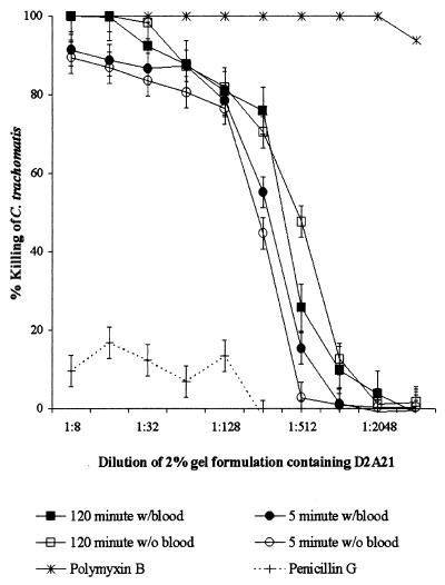
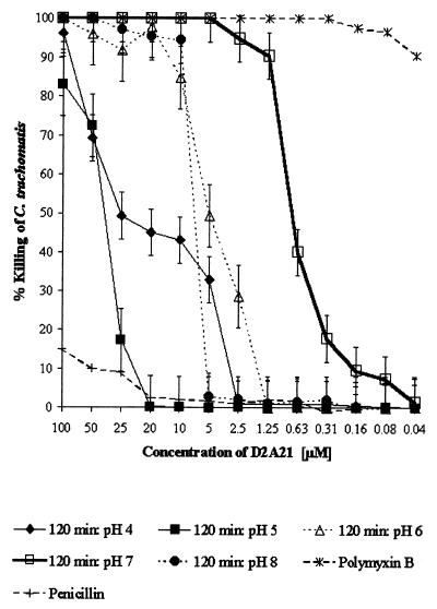
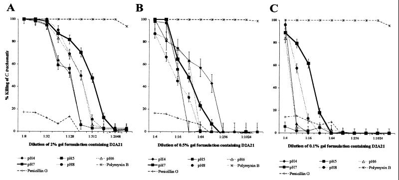
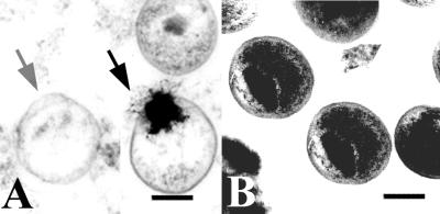
Topically applied microbicides that eradicate pathogens at the time of initial exposure represent a powerful strategy for the prevention of sexually transmitted infections. To aid in the further development of an effective topical microbicide, we assessed the minimum cidal concentration (MCC) of two cecropin peptides, D2A21 and D4E1, and gel formulations containing 0.1 to 2% D2A21 against Chlamydia trachomatis in vitro. The MCC of peptide D2A21 was 5 microM (18.32 microg/ml), and that of peptide D4E1 was 7.5 microM (21.69 microg/ml). The MCC of gel formulations containing 2% D2A21 was 0.2 mM (0.7 mg/ml), and that of gel formulations containing 0.5% D2A21 was 0.2 mM (0.7 mg/ml). There was no significant variation in the results when two different C. trachomatis strains were tested, and the addition of 10% human blood did not significantly alter the MCCs. pH values above and below 7 reduced the activity of the D2A21 peptide alone, but the MCC of the 2% D2A21 gel formulation was only slightly altered at the various pHs tested. Ultrastructural studies indicated that C. trachomatis membranes were disrupted after D2A21 exposure, resulting in leakage of the cytoplasmic contents. These in vitro results suggest that these cecropin peptides may be an effective topical microbicide against C. trachomatis and support the need for further evaluation.
Figures
References
-
- Bechinger, B. 1997. Structure and functions of channel-forming peptides: magainins, cecropins, melittin and alamethicin. J. Membr. Biol. 156:197–211. - PubMed
-
- Boisvert, J. F., L. A. Koutsky, R. J. Suchland, and W. E. Stamm. 1999. Clinical features of Chlamydia trachomatis rectal infection by serovar among homosexually active men. Sex. Transm. Dis. 26:392–398. - PubMed
-
- Boman, H. G. 1998. Gene-encoded peptide antibiotics and the concept of innate immunity: an update review. Scand J. Immunol 48:15–25. - PubMed
-
- Centers for Disease Control and Prevention. 1999. Summary of notifiable diseases, United States, 1998. Morb. Mortal. Wkly. Rep. 47:11–23. - PubMed
Publication types
MeSH terms
Substances
Grants and funding
LinkOut - more resources
Full Text Sources
Other Literature Sources
Medical
