. 2001 Dec 15;15(24):3308-18.
doi: 10.1101/gad.935501.

Mediator function of the human Rad51B-Rad51C complex in Rad51/RPA-catalyzed DNA strand exchange

Affiliations

Mediator function of the human Rad51B-Rad51C complex in Rad51/RPA-catalyzed DNA strand exchange

S Sigurdsson et al. Genes Dev. .

Abstract

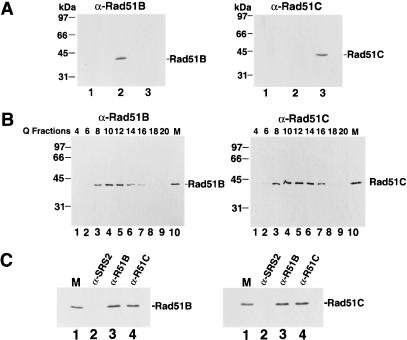
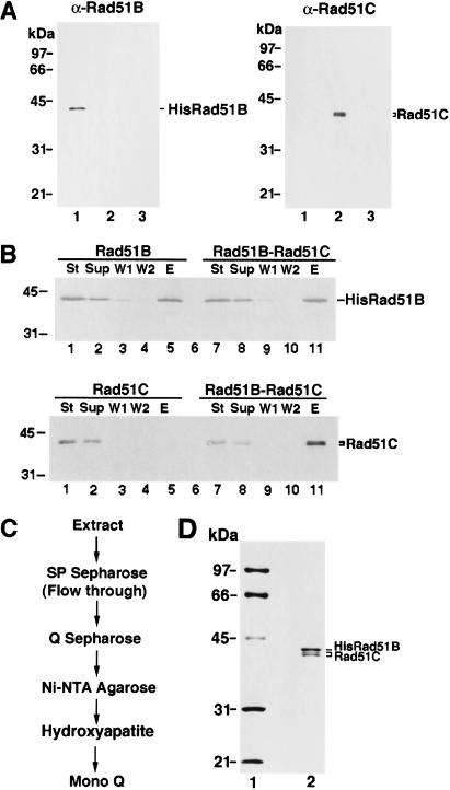
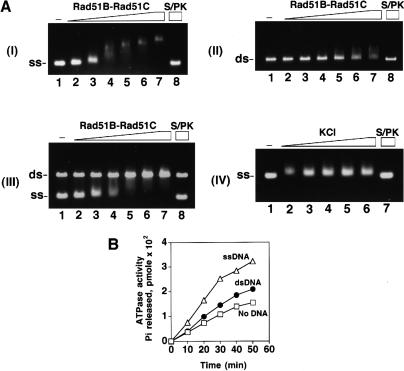
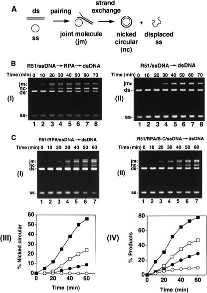
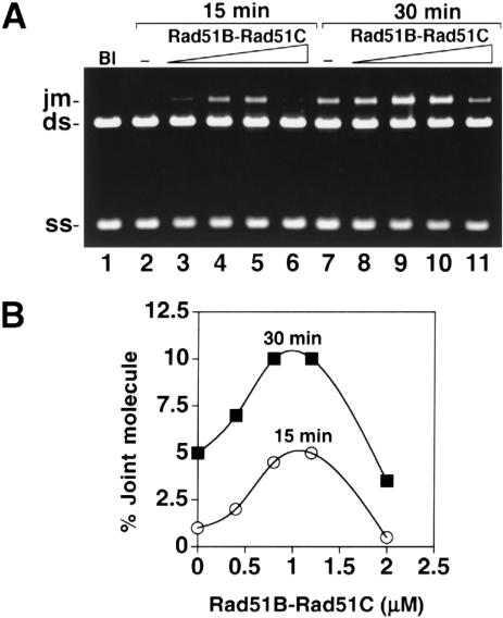
Five Rad51-like proteins, referred to as Rad51 paralogs, have been described in vertebrates. We show that two of them, Rad51B and Rad51C, are associated in a stable complex. Rad51B-Rad51C complex has ssDNA binding and ssDNA-stimulated ATPase activities. We also examined the functional interaction of Rad51B-Rad51C with Rad51 and RPA. Even though RPA enhances Rad51-catalyzed DNA joint formation via removal of secondary structure in the ssDNA substrate, it can also compete with Rad51 for binding to the substrate, leading to suppressed reaction efficiency. The competition by RPA for substrate binding can be partially alleviated by Rad51B-Rad51C. This recombination mediator function of Rad51B-Rad51C is likely required for the assembly of the Rad51-ssDNA nucleoprotein filament in vivo.


Figures

Figure 1
Figure 1
Rad51B and Rad51C form a stable complex. (A) Specificity of antibodies. Yeast cells harboring the empty expression vector pPM231 (2μ, GAL-PGK; lane 1), the Rad51B expression plasmid pR51B.1 (, GAL-PGK-RAD51B; lane 2), and the Rad51C expression plasmid pR51C.1 (2μ, PGK-RAD51C; lane 3) were run in an 11% polyacrylamide gel and then subjected to immunoblot analysis with either anti-Rad51B (αRad51B; left panel) or anti-Rad51C antibodies (αRad51C; right panel). (B) Rad51B and Rad51C are associated in a complex in human cells. HeLa cell extract was fractionated in a Q Sepharose column, and the indicated fractions were run in an 11% gel and then subjected to immunoblot analysis with anti-Rad51B antibodies (α-Rad51B; left panel) or anti-Rad51C antibodies (α-Rad51C; right panel). Yeast extracts containing Rad51B (lane 10 in left panel, marked M) or Rad51C (lane 10 in right panel, marked M) were used to help identify these proteins in the Q column fractions. (C) Coimmunoprecipitation of Rad51B and Rad51C from the Q column fractions. The Q Sepharose pool (fractions 8–16) was subjected to immunoprecipitation with protein A beads containing anti-ySrs2 antibodies (α-SRS2), anti-Rad51B antibodies (α-R51B) and anti-Rad51C antibodies (α-R51C). Proteins bound to the various immunobeads were eluted by SDS treatment and analyzed for their content of Rad51B (left panel) and Rad51C (right panel).
Figure 2
Figure 2
Purification of Rad51B and Rad51C from insect cells. (A) Expression of six-histidine-tagged Rad51B and Rad51C in insect cells. Nitrocellulose blots containing extracts from insect cells without any baculovirus (lane 3 of both panels) and infected with the recombinant 6His-tagged Rad51B baculovirus (lane 1 of both panels) or Rad51C baculovirus (lane 2 of both panels) were probed with either anti-Rad51B antibodies (αRad51B; left panel) or anti-Rad51C antibodies (αRad51C; right panel). (B) Complex formation between 6His-tagged Rad51B and Rad51C. Extracts from insect cells expressing 6His-tagged Rad51B (Rad51B), Rad51C (Rad51C), and the mixture of these extracts (Rad51B–Rad51C) were incubated with nickel-NTA agarose beads, which were washed with 10 mM, 20 mM, and then with 150 mM imadazole. The starting fractions (St), the supernatants containing unbound proteins (Sup), the 10 mM (W1) and 20 mM (W2) imidazole washes, and the 150 mM imidazole eluate (E) were subjected to immunoblotting to determine their content of Rad51B (upper panel) and Rad51C (lower panel). (C) Purification scheme for Rad51B–Rad51C complex. (D) Purity analysis. The purified Rad51B–Rad51C complex, 1.5 μg in lane 2, was run alongside molecular size markers (lane 1) in an 11% denaturing polyacrylamide gel and stained with Coomassie blue.
Figure 3
Figure 3
Rad51B–Rad51C binds DNA and hydrolyzes ATP. (A) Rad51B–Rad51C complex (0.15, 0.3, 0.45, 0.6, 0.75, and 0.9 μM in lanes 27, respectively) was incubated with φX ssDNA (12 μM nucleotides in panel I; designated as ss), φX dsDNA (4 μM base pair in panel II; designated as ds), or with both the ssDNA and dsDNA (panel III) for 10 min at 37°C and then run in a 0.9% agarose gel. The DNA species were stained with ethidium bromide. In lane 8 of all three panels, the nucleoprotein complex formed with 0.9 μM of Rad51B–Rad51C complex was treated with 0.5% SDS and 500 μg/mL proteinase K at 37°C for 5 min before loading onto the agarose gel. In lane 1 of all three panels, DNA was incubated in buffer without protein. In panel IV, Rad51B–Rad51C complex (0.3 μM) was incubated with ssDNA (12 μM nucleotides) in the presence of increasing concentrations (50, 100, 150, 200, and 250 mM in lanes 26, respectively) of KCl at 37°C for 10 min and then analyzed. (B) Rad51B–Rad51C, 1.8 μM, was incubated with 1 mM ATP in the absence of DNA (designated by the squares) and in the presence of ssDNA (20 μM nucleotides; designated by the triangles) or dsDNA (20 μM base pairs; designated by the closed circles) for the indicated times at 37°C.
Figure 4
Figure 4
Mediator function of Rad51B–Rad51C. (A) Schematic of the homologous DNA pairing and strand exchange reaction using φX174 DNA substrates. Linear duplex is paired with the homologous ssDNA circle to yield a joint molecule. DNA strand exchange, if successful over the length (5.4 kb) of the DNA molecules, results in the formation of the nicked circular duplex. (B) Rad51-mediated DNA pairing and strand exchange was carried out with RPA (panel I) or without it (panel II). In panel I, the ssDNA was preincubated with Rad51 (R51) before RPA was added. The concentrations of the reaction components were: Rad51, 7.5 μM; RPA, 1.5 μM; ssDNA, 30 μM nucleotides; linear duplex, 15 μM base pairs. (C) In the DNA strand exchange reaction in panel I, the ssDNA was incubated with both Rad51 (R51) and RPA simultaneously, and in the reaction in panel II, the ssDNA was incubated with Rad51, RPA and Rad51B–Rad51C (BC) simultaneously. The concentration of Rad51B–Rad51C was 0.8 μM, while the concentrations of the other components were exactly as those in B. In panel III, the amounts of nicked circular duplex in the reactions represented in B panel I (filled squares) and panel II (open circles) and in C panel I (filled circles) and panel II (open squares) are plotted. In panel IV, the amounts of total reaction products (sum of joint molecules and nicked circular duplex) in the reactions represented in B panel I (filled squares) and panel II (open circles) and in C panel I (filled circles) and panel II (open squares) are plotted.
Figure 5
Figure 5
Mediator activity as a function of Rad51B–Rad51C concentration. (A) The φX174 ssDNA template (30 μM nucleotides) was incubated with Rad51 (7.5 μM), RPA (1.5 μM), and increasing concentrations of Rad51B–Rad51C (0, 0.6, 0.8, 1.0, and 1.4 μM in lanes 15, respectively) for 10 min before the φX174 linear duplex (15 μM base pairs) was incorporated to complete the reaction mixtures. Portions of the reaction mixtures were withdrawn at 30 min (panel I), 60 min (panel II), and 80 min (panel III) and then processed for agarose gel electrophoresis. (B) The results from A and from two other independent experiments performed under the same reaction conditions were compiled and graphed. Symbols: results from the 30 min timepoint (squares), the 60 min timepoint (filled triangles), and the 80 min timepoint (circles). Panel I shows the levels of nicked circular duplex formed, and panel II shows the amounts of total reaction products (joint molecules and nicked circular duplex).
Figure 6
Figure 6
Rad51B–Rad51C stimulates DNA joint formation but does not replace RPA in DNA strand exchange. (A) Rad51 and Rad51B–Rad51C were used in the homologous DNA pairing and strand exchange reaction without RPA. The concentrations of the reaction components were: Rad51, 7.5 μM; Rad51B–Rad51C, 0.4 to 2 μM, as indicated; ssDNA, 30 μM nucleotides; linear duplex, 15 μM base pairs. (B) Graphic representation of the results from the experiment in A.

Similar articles

Cited by

References

    1. Baumann P, West SC. The human Rad51 protein: Polarity of strand transfer and stimulation by hRP-A. EMBO J. 1997;16:5198–5206. - PMC - PubMed
    1. ————— Heteroduplex formation by human Rad51 protein: Effects of DNA end-structure, hRP-A and hRad52. J Mol Biol. 1999;291:363–374. - PubMed
    1. Beernink HT, Morrical SW. RMPs: Recombination/replication mediator proteins. Trends Biochem Sci. 1999;24:385–389. - PubMed
    1. Benson FE, Stasiak A, West SC. Purification and characterization of the human Rad51 protein, an analogue of E. coli RecA. EMBO J. 1994;23:5764–5771. - PMC - PubMed
    1. Bianco PR, Tracy RB, Kowalczykowski SC. DNA strand exchange proteins: A biochemical and physical comparison. Front Biosci. 1998;3:D570–D603. - PubMed

Publication types