. 2001 Dec 15;15(24):3342-54.
doi: 10.1101/gad.946501.

Chromatin-specific regulation of LEF-1-beta-catenin transcription activation and inhibition in vitro

Affiliations

Chromatin-specific regulation of LEF-1-beta-catenin transcription activation and inhibition in vitro

A V Tutter et al. Genes Dev. .

Abstract

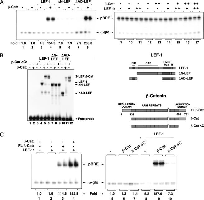
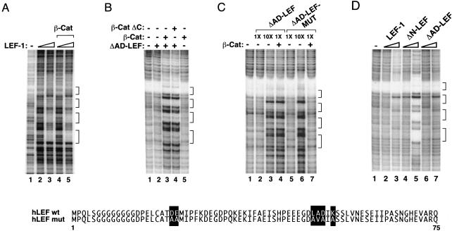
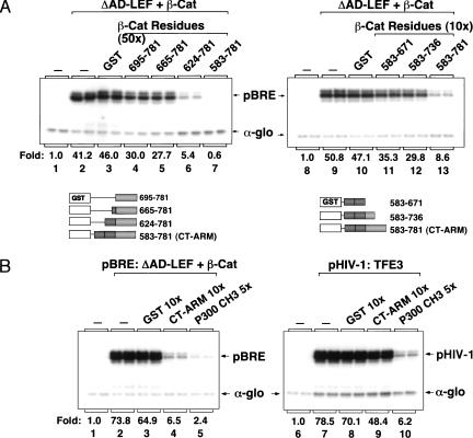
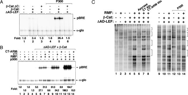
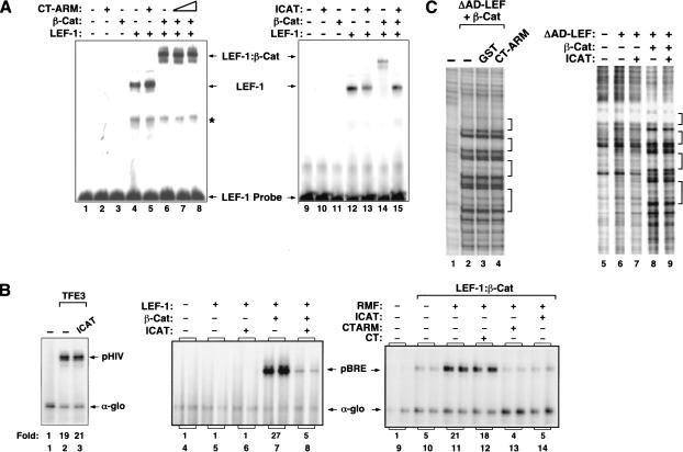
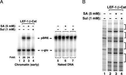
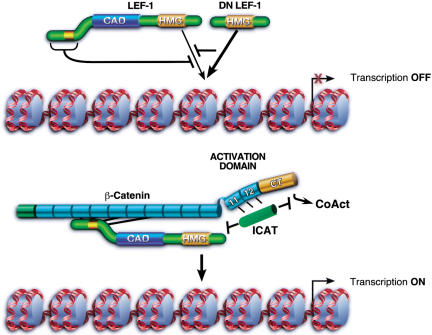
Transcriptional activation of Wnt/Wg-responsive genes requires the stabilization and nuclear accumulation of beta-catenin, a dedicated coactivator of LEF/TCF enhancer-binding proteins. Here we report that recombinant beta-catenin strongly enhances binding and transactivation by LEF-1 on chromatin templates in vitro. Interestingly, different LEF-1 isoforms vary in their ability to bind nucleosomal templates in the absence of beta-catenin, owing to N-terminal residues that repress binding to chromatin, but not nonchromatin, templates. Transcriptional activation in vitro requires both the armadillo (ARM) repeats and the C terminus of beta-catenin, whereas the phosphorylated N terminus is inhibitory to transcription. A fragment spanning the C terminus (CT) and ARM repeats 11 and 12 (CT-ARM), but not the CT alone, functions as a dominant negative inhibitor of LEF-1-beta-cat activity in vitro and can block ATP-dependent binding of the complex to chromatin. LEF-1-beta-cat transactivation in vitro was also repressed by inhibitor of beta-catenin and Tcf-4 (ICAT), a physiological inhibitor of Wnt/Wg signaling that interacts with ARM repeats 11 and 12, and by the nonsteroidal anti-inflammatory compound, sulindac. None of these transcription inhibitors (CT-ARM, ICAT, or sulindac) could disrupt the LEF-1-beta-cat complex after it was stably bound to chromatin. We conclude that the CT-ARM region of beta-catenin functions as a chromatin-specific activation domain, and that several inhibitors of the Wnt/Wg pathway directly modulate LEF-1-beta-cat activity on chromatin.


Figures

Figure 1
Figure 1
Chromatin-specific activation of transcription by purified recombinant LEF-1 and β-catenin. (A) Primer-extension analysis of pBRE transcription in the presence of wild-type or mutant LEF-1 proteins on chromatin (left panel) or nonchromatin (right panel) templates in vitro. Reactions either lacked enhancer factors (lane 1), or contained β-cat (120 nM, lanes 2,4,6,8), full-length LEF-1 (120 nM, lanes 3,4), ΔN-LEF (120 nM, lanes 5,6), or ΔAD-LEF (120 nM, lanes 7,8). (Right panel) Transcription reactions either lacked enhancer factors (lane 9), or contained LEF-1 (250 nM, lanes 10,14,16; 750 nM, lanes 11,15,17), β-cat (250 nM, lanes 12,14,15; 750 nM, lanes 13,16,17). Arrows designate transcription from the pBRE template (pBRE) or the alpha-globin promoter (α-glo), which was added as a nonchromatin template to the HeLa transcription extract as a positive control for RNA recovery. (B) pBRE chromatin transcription reactions with full-length (FLβ-cat) and truncated β-cat proteins (β-cat and β-catΔC). Reactions either lacked enhancer factors (lanes 1,5) or contained LEF-1 (120 nM, lanes 2–4 and 8–10), FLβ-cat (120 nM, lane 3), β-cat (120nM, lanes 4,6,9), or β-cat ΔC (120 nM, lanes 7,10). (C) Western blot analysis of β-cat and FLβ-cat proteins before (t = 0) and after (t = 5 h) chromatin assembly. At the right is a schematic of the different mutant LEF-1 and β-catenin proteins examined in this study.
Figure 2
Figure 2
β-catenin strongly enhances the binding of LEF-1 to chromatin, but not nonchromatin templates. (A) DNase I footprint analysis of binding of LEF-1 and LEF-1–β-cat to (nonchromatin) pBRE DNA. Binding reactions either lacked enhancer factors (lane 1) or contained LEF-1 (112 nM, lanes 2,4; 560 nM, lanes 3,5) and β-cat (560 nM, lanes 4,5). The four LEF-1 binding sites in the pBRE enhancer are indicated with brackets. (B) DNase I footprint analysis of pBRE chromatin assembled in the absence of enhancer factors (lane 1) or in the presence of ΔAD-LEF (120 nM, lanes 2–4), β-cat (120 nM, lanes 3,5) or β-cat ΔC (120 nM, lane 4). (C) DNase I footprint analysis of pBRE chromatin assembled in the absence of enhancer factors (lane 1) or in the presence of β-cat (120 nM, lanes 4,7), ΔAD-LEF (112 nM, lanes 2,4; 1 μM, lane 3), or ΔAD-LEF-MUT (112 nM, lanes 5,7; 1 μM, lane 6). Substitutions in the amino terminus of ΔAD-LEF-MUT that abrogate binding to β-cat are indicated with bold type. (D) The N terminus of LEF-1 inhibits binding to chromatin, but not nonchromatin, templates. DNase I footprint analysis of the binding of LEF-1 and mutant LEF-1 proteins to pBRE chromatin. Chromatin assembly reactions either lacked enhancer factors (lane 1) or contained full-length LEF-1 (120 nM, lane 2; 1 μM, lane 3), ΔN-LEF (120 nM, lane 4; 1 μM, lane 5), or ΔAD-LEF (120 nM, lane 6; 1 μM, lane 7). Brackets indicate the four LEF/TCF binding sites in the pBRE enhancer.
Figure 3
Figure 3
The β-cat CT–ARM domain fragment selectively inhibits LEF-1–β-cat transcription on chromatin in vitro. (A) (Left panel) Chromatin was assembled in absence of enhancer factors (lane 1) or with 120 nM ΔAD-LEF and 120 nM β-cat (lanes 2–7) in the presence of a 50-fold molar excess (relative to β-cat) of either GST alone (lane 3), GST–β-cat aa695–781 (lane 4), GST–β-cat aa665–781 (lane 5), GST–β-cat aa624–781 (lane 6), or GST–β-cat aa583–781 (CT–ARM, lane 7). (Right panel) Chromatin was assembled in the absence of enhancer factors (lane 8) or with 120 nM ΔAD-LEF and 120 nM β-cat (lanes 9–13) and a 10-fold molar excess (relative to β-cat) of either GST alone (lane 10), GST–β-cat amino acids 583–671 (lane 11), GST–β-cat amino acids 583–736 (lane 12), or GST–β-cat amino acids 583–781 (lane 13). The GST–β-cat fragments are represented schematically below the figure. (B) Comparison of the transcription inhibitory effects of the β-cat CT–ARM and p300 CH3 domains on pBRE (left panel) and HIV-1 (right panel) chromatin templates. (Left panel) Transcription reactions either lacked enhancer factors (lane 1) or contained 120 nM ΔAD-LEF and 120 nM β-cat (lanes 2–5), along with a 10-foldmolar excess (relative to β-cat) of GST (lane 3), GST–CT–ARM (lane 4) or the GST–p300/CH3 fragment (lane 5). (Right panel) Transcription reactions either lacked enhancer factors (lane 6) or contained GST–TFE3 (30 nM, lanes 7–10) and a 10-fold molar excess (relative to TFE3) of GST (lane 8), GST–CT–ARM (lane 9) or the GST–p300/CH3 fragment (lane 10). Arrows indicate pBRE, pHIV-1, and α-globin transcripts.
Figure 4
Figure 4
p300 and RMF enhance transcription from fully assembled chromatin templates by LEF-1 and β-catenin in vitro. (A) Chromatin transcription reactions either lacked enhancer factors (lane 1), or contained ΔAD-LEF (120 nM, lanes 2,3,5,6), β-cat (120 nM, lanes 2,5), or β-cat ΔC (120 nM, lanes 3,6). Where indicated, purified recombinant p300 (60 nM, lanes 4–6) was incubated together with LEF-1 and β-cat after completion of pBRE chromatin assembly. (B) Transcription reactions either lacked enhancer factors (lane 1), or contained 120 nM ΔAD-LEF and 120 nM β-cat (lanes 5–14) added after pBRE chromatin assembly. Where indicated, reactions also contained p300 (60 nM, lanes 3,4,9–14), RMF (1 μg, lanes 2,4,6–8,12–14), GST–CT (4.2 μg, lanes 7,10,13), or GST–CT–ARM (4.8 μg, lanes 8,11,14), added simultaneously with LEF-1 and β-cat. (C) Binding of LEF-1–β-cat to assembled pBRE chromatin templates in the presence or absence of p300, RMF, and the CT–ARM inhibitor. Binding reactions either lacked enhancer factors (lanes 1,9) or contained ΔAD-LEF (120 nM, lanes 3–8,10–11,13–14), β-cat (120 nM, lanes 4–8,10–11,13–14), p300 (60 nM, lanes 12–14), RMF (1 μg, lanes 2,5–8,11,14), or a 10-fold excess of GST–CT (lane 7), GST–CT–ARM (lane 8), and apyrase (0.5 unit; lane 6), added with the LEF-1–β-cat complex after chromatin assembly.
Figure 5
Figure 5
ICAT is a potent and selective inhibitor of LEF-1–β-cat transcription on chromatin. (A) Analysis of the effects of CT–ARM and ICAT on the formation of the LEF-1–β-cat–DNA ternary complex in gel mobility shift experiments. Binding reactions either lacked enhancer factors (lanes 1,9) or contained 110 nM LEF-1 (lanes 4–8,12–15), 110 nM β-cat (lanes 3,6–8,11,14,15), GST–CT–ARM (500 nM, lane 7; 2.5 μM, lanes 2,5,8) or GST–ICAT (2.5 μM, lanes 10,13,15). Arrows indicate the position of LEF-1–DNA and LEF-1–β-cat–DNA complexes. Asterisk indicates nonspecific band. (B) Analysis of the effect of ICAT on HIV-1 (lanes 1–3) and pBRE transcription (lanes 4–14) in vitro. Chromatin was assembled in the absence of enhancer factors (lanes 1,4,9) or in the presence of 120 nM GST–TFE3 (lanes 2,3), or 120 nm ΔAD-LEF and 120 nM β-cat (lanes 5–8,10–14). Where indicated, reactions also contained 1.2 μM of ICAT (lanes 3,6,8,14). The enhancer factors were either incubated with the template during chromatin assembly (lanes 4–8) or incubated after chromatin assembly together with 1.0 μg RMF (lanes 11–14). Arrows indicate the pHIV, pBRE, and α-globin transcripts. (C) DNase I footprint analysis of the effects of CT–ARM and ICAT on the β-catenin-enhanced binding of LEF-1 to chromatin. Binding reactions either lacked enhancer factors (lanes 1,5) or contained 120 nM ΔAD-LEF and 120 nM β-cat (lanes 2–4,6,7). Where indicated, reactions also contained 1 μM each of GST (lane 3), GST–CT–ARM (lane 4), or GST–ICAT (lane 7).
Figure 6
Figure 6
LEF-1–β-cat transactivation on chromatin in vitro is inhibited by the NSAID, sulindac. (A) (Left panel) Analysis of the effects of NSAID on LEF-1–β-cat transcription of pBRE chromatin templates in vitro. Chromatin was assembled in the absence of enhancer factors (lane 1) or in the presence of 120 nM ΔAD-LEF and 120 nM β-cat (lanes 2–4), in the presence of 5 mM salicylic acid (lane 3) or 1 mM sulindac (lane 4). (Right panel) Analysis of the effects of NSAIDs on nonchromatin pBRE DNA templates. Reactions either lacked NSAIDs (lane 5) or contained either 5 mM salicylic acid (lane 6) or 1 mM sulindac (lane 7). Arrows indicate pBRE (chromatin) and α-globin (nonchromatin) transcripts. (B) DNase I footprint analysis of the effects of NSAIDs on binding of the LEF-1–β-cat complex to chromatin. Chromatin was assembled in the absence of enhancer factors (lane 1) or in the presence of 120 nM each of ΔAD-LEF and β-cat (lanes 2–4), incubated with either 5 mM salicylic acid (lane 3) or 1 mM sulindac (lane 4). Brackets indicate the four LEF/TCF binding sites in the pBRE enhancer.
Figure 7
Figure 7
Chromatin-specfic effects of β-catenin on binding and transcriptional activation by LEF-1 in vitro. In the absence of β-catenin, Wnt-responsive genes are repressed by LEF/TCF–Groucho complexes, and binding of uncomplexed LEF-1 to chromatin is inhibited through amino-terminal residues. LEF-1 isoforms that lack the inhibitory amino-terminal domain bind chromatin more avidly and function as potent feedback inhibitors of Wnt signaling in vivo (dominant negative DN LEF-1). The interaction of the central ARM repeats of β-catenin with the N terminus of LEF-1 alleviates this inhibition, and LEF-1 binds cooperatively with its coactivator to chromatin. Optimal transactivation requires an activation domain located at the C terminus that may also include C-terminal ARM repeats 11 and 12. LEF-1–β-cat transcription can be selectively blocked in vitro by a dominant-negative fragment of β-catenin that spans the carboxyl terminus and ARM repeats 11/12, as well as by the physiological Wnt inhibitor, ICAT. ICAT, but not CT–ARM, can disrupt the interaction between LEF-1 and β-catenin. Moreover, ICAT can block transcription from complexes bound stably to chromatin, indicating that it may also disrupt interactions between β-catenin and downstream transcriptional coactivators (CoAct).

Similar articles

Cited by

References

    1. Aoki M, Hecht A, Kruse U, Kemler R, Vogt PK. Nuclear endpoint of Wnt signaling: Neoplastic transformation induced by transactivating lymphoid-enhancing factor 1. Proc Natl Acad Sci. 1999;96:139–144. - PMC - PubMed
    1. Armstrong JA, Bieker JJ, Emerson BM. A SWI/SNF-related chromatin remodeling complex, E-RC1, is required for tissue-specific transcriptional regulation by EKLF in vitro. Cell. 1998;95:93–104. - PubMed
    1. Barker N, Hurlstone A, Musisi H, Miles A, Bienz M, Clevers H. The chromatin remodeling factor Brg-1 interacts with β-catenin to promote target gene activation. EMBO J. 2001;20:4935–4943. - PMC - PubMed
    1. Bauer A, Huber O, Kemler R. Pontin52, an interaction partner of beta-catenin, binds to the TATA box binding protein. Proc Natl Acad Sci. 1998;95:14787–14792. - PMC - PubMed
    1. Behrens J, von Kries JP, Kuhl M, Bruhn L, Wedlich D, Grosschedl R, Birchmeier W. Functional interaction of beta-catenin with the transcription factor LEF-1. Nature. 1996;382:638–642. - PubMed

Publication types

MeSH terms