Comparative Study
. 2001 Dec 15;15(24):3355-64.
doi: 10.1101/gad.931001.

A homeobox gene, PRESSED FLOWER, regulates lateral axis-dependent development of Arabidopsis flowers

Affiliations
Comparative Study

A homeobox gene, PRESSED FLOWER, regulates lateral axis-dependent development of Arabidopsis flowers

N Matsumoto et al. Genes Dev. .

Erratum in

  • Genes Dev 2002 Mar 15;16(6):764

Abstract

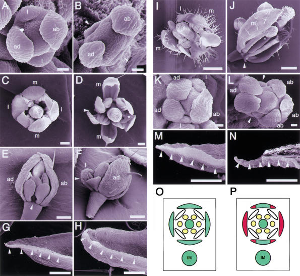
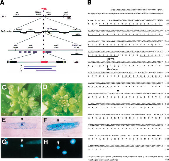
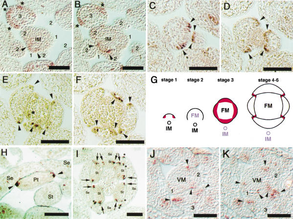
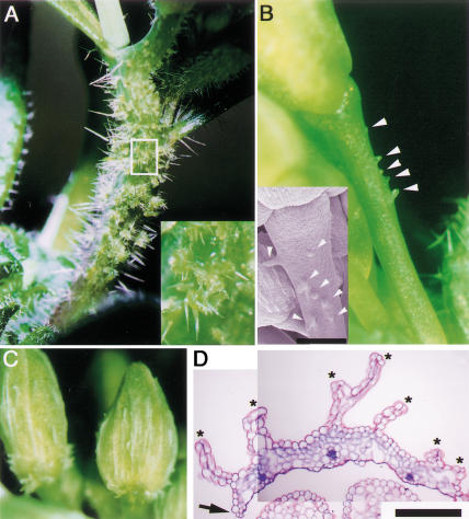
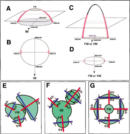
It is postulated that the symmetric organization of plant lateral organs is based on two crossed axes, the abaxial-adaxial and the lateral axes. The PRESSED FLOWER (PRS) gene, the expression and function of which are dependent on the lateral axis, is reported in this study. In the prs mutant, growth of the lateral sepals is repressed, and although the size and shape of the abaxial and adaxial sepals are normal, the cell files at the lateral margins are missing. Double-mutant analyses showed that the PRS gene functions independently of the determinations of both floral organ identity and floral meristem size. The PRS gene, encoding a putative transcriptional factor with a homeodomain, was shown to be required for cell proliferation. PRS gene expression is spatially and temporally unique and is expressed in a restricted number of L1 cells at the lateral regions of flower primordia, floral organ primordia, and young leaf primordia. Our study strongly suggests that the PRS gene is involved in the molecular mechanism of lateral axis-dependent development of lateral organs in Arabidopsis.


Figures

Figure 1
Figure 1
Phenotypes of mutant flowers. (A) A wild-type flower at stage 4. (B) A prs flower at stage 4. Lateral sepal primordia are not observed (arrowheads). (C) A mature flower of wild type. (D) A mature flower of prs. One lateral sepal is missing (arrowhead), and the other is smaller than normal (asterisk). (E) A young flower of prs. A lateral sepal is completely absent (arrowhead). (F) A young flower of prs. A lateral sepal (arrowhead) is smaller than the adaxial sepal. (G) Knife-edge margin of a wild-type sepal (arrowheads). (H) Margin of a prs sepal. The knife-edge cells are absent (arrowheads). (I) An ap2-1 flower. (J) A prs ap2-1 flower. A transformed leaf-like organ at lateral position remained as a filamentous organ (arrowhead). (K) A clv1-4 flower. (L) A prs clv1-4 flower. Sepal primordia are absent at the lateral regions (arrowheads). (M) Margin of an ap3-5 sepal in the second whorl. The knife-edge cells are shown by arrowheads. (N) Margin of a prs ap3-5 sepal in the second whorl. The knife-edge cells are absent (arrowheads). (O) Diagram of a wild-type flower. (P) Diagram showing positions of defects observed in a prs flower (shown in red). (ab) abaxial; (ad) adaxial; (l) lateral; (m) medial; (IM) inflorescence meristem. A,B,K,L, bar, 20 μm; CH,M,N, bar, 200 μm; I,J, bar, 500 μm.
Figure 2
Figure 2
Scheme of the positional cloning and the structure of PRS. (A) Genomic structure of the PRS (T8O18.10) region on chromosome 2. PRS was mapped between markers nmB and nmG. Numbers below the markers indicate the recombination frequency between the marker and the PRS locus. The red bar and arrow show the predicted ORF of T8O18.10. Blue lines show the genomic clones that complemented the prs phenotype. (B) cDNA sequence of PRS and the predicted amino acid sequence including a homeodomain (double underline), a glutamine-rich region (single underline), and a histidine-rich region (broken line). Mutation site in prs is shown. The position of the intron (437 bp) is indicated by a triangle (GenBank accession no. AB058920). (C) Inflorescence of prs. (D) Inflorescence of prs transformed with a genomic clone including T8O18.10. (EH) Subcellular localization of the PRS-GUS fusion protein by particle bombardment of onion epidermal cells. (E,G) Transiently transformed with 35S:PRS-GUS. (F,H) Transiently transformed with 35S:GUS as a control. GUS (E,F) and DAPI (G,H) staining are shown. Arrowheads indicate the nucleus.
Figure 3
Figure 3
Expression patterns of PRS in inflorescence, floral organs, and leaf primordia of wild type. (A,B) PRS expression patterns in an inflorescence in serial sections. Numbers show the stage of flowers. Arrowheads and asterisks show expressions at early stage 1 and at stage 3, respectively. (CF) PRS expression patterns in sepals in serial sections at stage 5. Arrowheads show the expressing cells at the lateral edges of sepal primordia. In E, the asterisk shows the expression in a petal primordium. (G) Schematic drawings that show the PRS expression patterns. At early stage 1, the PRS expression is restricted at the lateral regions of a flower primordium. At stage 2, the PRS expression is disappeared. At stage 3, the PRS expression is reappeared in all four-sepal young primordia. After stage 4, the PRS expression is restricted at the lateral edges of young sepals. (H) Expression of PRS at the lateral edges of a petal (arrowheads). (I) Expression of PRS at the lateral edges of stamens (arrows) and of sepals (asterisks). No expression is detected in the carpel. (J,K) PRS-expression patterns in a vegetative meristem in serial sections. Numbers show the leaf primordium in order. Arrowheads show the expressions at the lateral edges of primordia. (IM) Inflorescence meristem; (FM) floral meristem; (VM) vegetative meristem; (Se) sepal; (Pt) petal; (St) stamen. Bars, 50 μm.
Figure 4
Figure 4
Phenotypes of 35S:PRS transgenic plants. (A) Multicellular bulges on the stem. (Inset) Magnification of the white box. (B) Multicellular bulges on the peduncle (arrowheads). (Inset) Magnified picture. (C) White wrinkle structures on sepals. (D) Transverse section of a sepal. The arrow and asterisks indicate the edge of the sepal and the wrinkle structures, respectively. B inset, bar, 250 μm; D, bar, 100 μm.
Figure 5
Figure 5
Diagrams of PRS expression and axes in lateral organs and in the flower. Diagrams showing the expression patterns of PRS in a flower primordium at stage 1 (A, B), and in a floral organ or a leaf (C, D). Red color indicates the position of cells expressing PRS. (E) Top view of a vegetative shoot showing abaxial–adaxial and lateral axes in leaf primordia. (F) Top view of an inflorescence showing abaxial–adaxial and lateral axes in early stage flowers. (G) Abaxial–adaxial and lateral axes in floral organs. (Ab-Ad) Abaxial–adaxial axis; (L-L) lateral axis; (VM) vegetative meristem; (IM) inflorescence meristem; (FM) floral meristem.

Similar articles

Cited by

References

    1. Abe M, Takahashi T, Komeda Y. Identification of a cis-regulatory element for L1 layer-specific gene expression, which is targeted by an L1-specific homeodomain protein. Plant J. 2001;26:487–494. - PubMed
    1. Bossinger G, Smyth DR. Initiation patterns of flower and floral organ development in Arabidopsis thaliana. Development. 1996;122:1093–1102. - PubMed
    1. Bowman JL. Morphology of the expanded first leaves. In: Bowman JL, editor. Arabidopsis: An atlas of morphology and development. New York, NY: Springer-Verlag; 1994. pp. 38–39.
    1. Bowman JL, Smyth DR, Meyerowitz EM. Genes directing flower development in Arabidopsis. Plant Cell. 1989;1:37–52. - PMC - PubMed
    1. ————— Genetic interactions among floral homeotic genes of Arabidopsis. Development. 1991;112:1–20. - PubMed

Publication types

MeSH terms