. 2002 Jan;184(2):459-67.
doi: 10.1128/JB.184.2.459-467.2002.

Bacillus subtilis tolerance of moderate concentrations of rifampin involves the sigma(B)-dependent general and multiple stress response

Affiliations

Bacillus subtilis tolerance of moderate concentrations of rifampin involves the sigma(B)-dependent general and multiple stress response

Julia Elisabeth Bandow et al. J Bacteriol. 2002 Jan.

Abstract

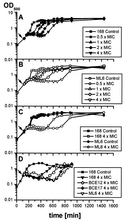
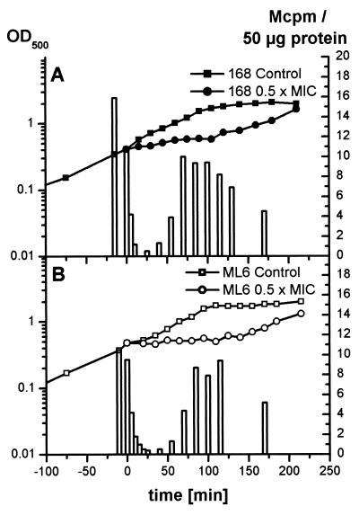
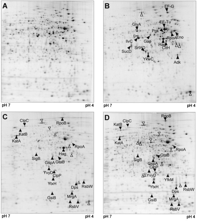
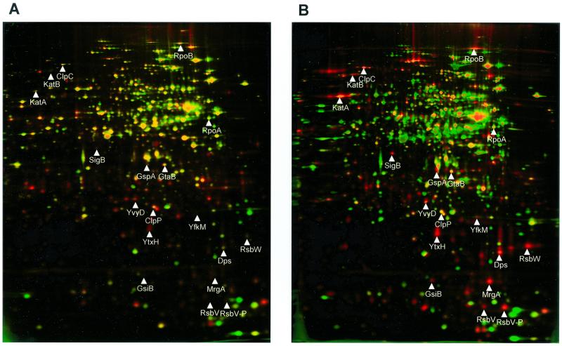
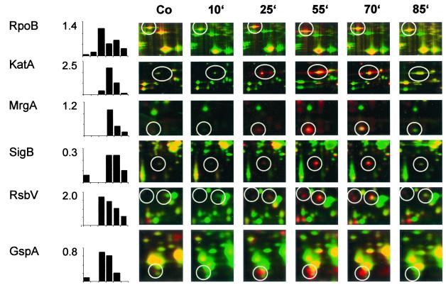
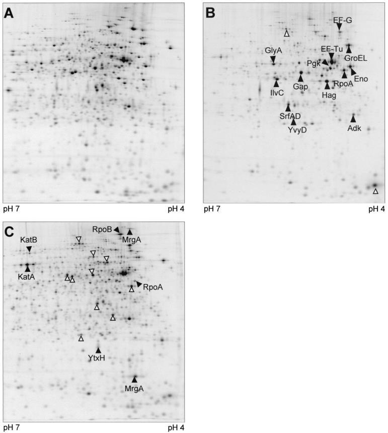
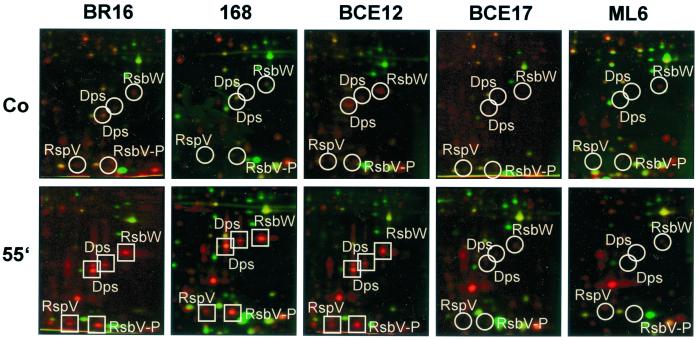
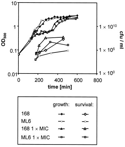
Low concentrations of the RNA polymerase inhibitor rifampin added to an exponentially growing culture of Bacillus subtilis led to an instant inhibition of growth. Survival experiments revealed that during the growth arrest the cells became tolerant to the antibiotic and the culture was able to resume growth some time after rifampin treatment. L-[(35)S]methionine pulse-labeled protein extracts were separated by two-dimensional polyacrylamide gel electrophoresis to investigate the change in the protein synthesis pattern in response to rifampin. The sigma(B)-dependent general stress proteins were found to be induced after treatment with the antibiotic. Part of the oxidative stress signature was induced as indicated by the catalase KatA and MrgA. The target protein of rifampin, the beta subunit (RpoB) of the DNA-dependent RNA polymerase, and the flagellin protein Hag belonging to the sigma(D) regulon were also induced. The rifampin-triggered growth arrest was extended in a sigB mutant in comparison to the wild-type strain, and the higher the concentration, the more pronounced this effect was. Activity of the RsbP energy-signaling phosphatase in the sigma(B) signal transduction network was also important for this protection against rifampin, but the RsbU environmental signaling phosphatase was not required. The sigB mutant strain was less capable of growing on rifampin-containing agar plates. When plated from a culture that had already reached stationary phase without previous exposure to the antibiotic during growth, the survival rate of the wild type exceeded that of the sigB mutant by a factor of 100. We conclude that the general stress response of B. subtilis is induced by rifampin depending on RsbP activity and that loss of SigB function causes increased sensitivity to the antibiotic.


Figures

FIG. 1.
FIG. 1.
Northern blot analysis of B. subtilis 168 extracts of control cells (lanes Co) and cultures challenged with rifampin for 4 (lanes 1) and 55 (lanes 2) min were performed with the digoxigenin-labeled RNA probes tufA (1.2 kb, tufA monocistronic; 5.5 kb, ybxF-rpsL-rpsG-fusA-tufA transcript) (A), sigB (2.2 kb, rsbV-rsbW-sigB-rsbX transcript) (B), and gspA (0.9 kb, gspA monocistronic) (C).
FIG. 2.
FIG. 2.
B. subtilis 168 (A), ML6 (sigB) (B), BCE12 (rsbU) (D), and BCE17 (rsbP) (D) were grown in synthetic medium to an OD500 of 0.4. The arrow indicates the time of addition of different concentrations of rifampin to the culture; the control was left untreated. (C) Comparison of the curves of B. subtilis 168 and ML6 treated with 0.24 μg of rifampin and untreated controls in one diagram.
FIG. 3.
FIG. 3.
The incorporation of l-[35S]methionine into 50 μg of B. subtilis 168 (A) and ML6 (sigB) (B) protein during 5 min of pulse-labeling was monitored parallel to the growth curve. The control sample was labeled immediately before rifampin application. Rifampin (0.03 μg/ml) was added at time zero.
FIG. 4.
FIG. 4.
Autoradiographs of 2D gels, pH 4 to 7, of B. subtilis 168 l-[35S]methionine-labeled protein extracts. (A) Control; (B to D) 10 to 15 min, 40 to 45 min, and 70 to 75 min, respectively, after treatment with rifampin at 0.03 μg/ml. Arrowheads indicate proteins with increased relative synthesis rates; protein spots marked by empty arrows have not been identified.
FIG. 5.
FIG. 5.
Dual-channel images of 2D gels (4) produced with Delta2D Software (DECODON GmbH) illustrate the change in protein synthesis (red) and protein accumulation (green). B. subtilis 168 control extract (A); 55 min after rifampin application (B).
FIG. 6.
FIG. 6.
Details of dual-channel images of 2D gels are shown to illustrate the change in B. subtilis 168 protein synthesis (red) and protein accumulation (green) at various times (labels at top) after rifampin application. Percentages of total protein synthesis generated with the Delta2D Software (DECODON GmbH) are shown for proteins, including the target protein RpoB, proteins of the oxidative stress signature (KatA and the monomer of MrgA), and the ςB-dependent proteins SigB, RsbV, and GspA. Co, control.
FIG. 7.
FIG. 7.
Autoradiographs of 2D gels, pH 4 to 7, of B. subtilis ML6 (sigB) l-[35S]methionine-labeled protein extracts. (A) control; (B and C) 10 to 15 min and 70 to 75 min, respectively, after application of rifampin (0.03 μg/ml). Solid arrowheads indicate proteins with increased relative synthesis rates; protein spots marked by empty arrows have not been identified.
FIG. 8.
FIG. 8.
Details of dual-channel images of 2D gels illustrate the difference in protein synthesis (red) and protein accumulation (green) after rifampin application among B. subtilis BR16, 168, BCE12 (rsbU), BCE17 (rsbP), and ML6 (sigB). The positions of induced proteins (squares) and the expected positions of proteins (circles) are indicated. Abbreviations: Co, control; 55′, 55 min.
FIG. 9.
FIG. 9.
Growth of B. subtilis 168 and ML6 (sigB) was monitored by measuring OD500. Both strains were challenged with rifampin (0.06 μg/ml) at an OD500 of 0.4. Aliquots of the cultures were plated on rifampin agar plates containing rifampin (0.06 μg/ml) to test the level of tolerance to the antibiotic.

References

    1. Anagnostopoulos, C., and J. Spizizen. 1961. Requirements for transformation in Bacillus subtilis. J. Bacteriol. 81:741–746. - PMC - PubMed
    1. Antelmann, H., S. Engelmann, R. Schmid, A. Sorokin, A. Lapidus, and M. Hecker. 1997. Expression of a stress- and starvation-induced dps/pexB-homologous gene is controlled by the alternative sigma factor ςB in Bacillus subtilis. J. Bacteriol. 179:7251–7256. - PMC - PubMed
    1. Antelmann, H., J. Bernhardt, R. Schmid, and M. Hecker. 1995. A gene at 333 degrees on the Bacillus subtilis chromosome encodes the newly identified SigB-dependent general stress protein GspA. J. Bacteriol. 177:3540–3545. - PMC - PubMed
    1. Bernhardt, J., K. Büttner, C. Scharf, and M. Hecker. 1999. Dual channel imaging of two-dimensional electropherograms in Bacillus subtilis. Electrophoresis 20:2225–2240. - PubMed
    1. Campbell, E. A., N. Korzheva, A. Mustaev, K. Murakami, S. Nair, A. Goldfarb, and S. A. Darst. 2001. Structural mechanism for rifampicin inhibition of bacterial RNA polymerase. Cell 104:901–912. - PubMed

Publication types

MeSH terms

LinkOut - more resources