

Novel form of fibronectin from zebrafish mediates infectious hematopoietic necrosis virus infection
- PMID: 11752139
- PMCID: PMC136842
- DOI: 10.1128/jvi.76.2.492-498.2002
Novel form of fibronectin from zebrafish mediates infectious hematopoietic necrosis virus infection
Abstract
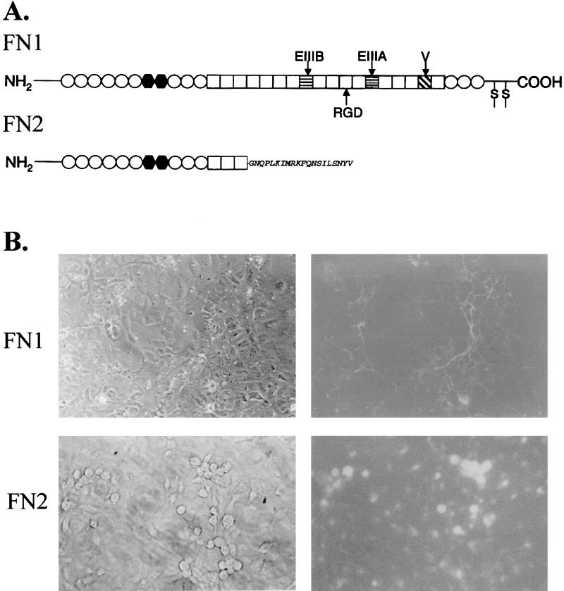
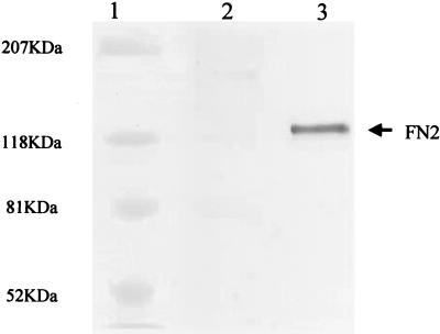
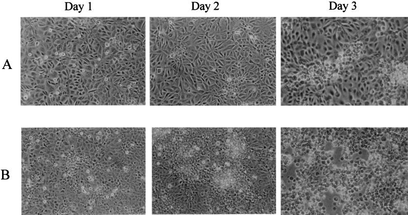
The presence of a novel form of zebrafish fibronectin (FN2) on the cell surface increased the cell's susceptibility to infection by infectious hematopoietic necrosis virus (IHNV). Unlike other fibronectins, FN2 possesses a truncated structure and accumulates on the cell surface instead of in the extracellular matrix. Fish embryo cells expressing recombinant FN2 were more susceptible to IHNV infection, with a greater percentage of cells exhibiting cytopathic effect (CPE) compared to nontransfected control cells. Incubation of nontransfected cells with soluble recombinant FN2 increased IHNV infection, as measured by plaque assay. The number of plaques increased in correlation with the amount of protein added and the length of time that cells were incubated with the protein. Incubation of IHNV with soluble FN2 before addition to cells also increased infection. FN2 immobilized on the culture surface inhibited IHNV infection. The results indicate that FN2 present on the cell surface is able to mediate IHNV attachment and cell entry.
Figures
Similar articles
-
The zebrafish galectins Drgal1-L2 and Drgal3-L1 bind in vitro to the infectious hematopoietic necrosis virus (IHNV) glycoprotein and reduce viral adhesion to fish epithelial cells.Dev Comp Immunol. 2016 Feb;55:241-252. doi: 10.1016/j.dci.2015.09.007. Epub 2015 Sep 30. Dev Comp Immunol. 2016. PMID: 26429411 Free PMC article.
-
Structure of the zebrafish galectin-1-L2 and model of its interaction with the infectious hematopoietic necrosis virus (IHNV) envelope glycoprotein.Glycobiology. 2019 May 1;29(5):419-430. doi: 10.1093/glycob/cwz015. Glycobiology. 2019. PMID: 30834446 Free PMC article.
-
In vitro effects of recombinant zebrafish IFN on spring viremia of carp virus and infectious hematopoietic necrosis virus.J Interferon Cytokine Res. 2006 Apr;26(4):256-9. doi: 10.1089/jir.2006.26.256. J Interferon Cytokine Res. 2006. PMID: 16704302
-
Identification and characterization of a novel fibronectin in zebrafish.Exp Cell Res. 2001 Aug 15;268(2):211-9. doi: 10.1006/excr.2001.5291. Exp Cell Res. 2001. PMID: 11478847
-
Identification of amino acid residues in infectious hematopoietic necrosis virus (IHNV) NV protein necessary for viral replication and pathogenicity.Fish Shellfish Immunol. 2018 Aug;79:294-302. doi: 10.1016/j.fsi.2018.05.032. Epub 2018 May 18. Fish Shellfish Immunol. 2018. PMID: 29782916
Cited by
-
Zebrafish as a Model for Fish Diseases in Aquaculture.Pathogens. 2020 Jul 27;9(8):609. doi: 10.3390/pathogens9080609. Pathogens. 2020. PMID: 32726918 Free PMC article. Review.
-
Susceptibilities of medaka (Oryzias latipes) cell lines to a betanodavirus.Virol J. 2010 Jul 12;7:150. doi: 10.1186/1743-422X-7-150. Virol J. 2010. PMID: 20624282 Free PMC article.
-
Zebrafish encoded 3-O-sulfotransferase-2 generated heparan sulfate serves as a receptor during HSV-1 entry and spread.Biochem Biophys Res Commun. 2013 Mar 22;432(4):672-6. doi: 10.1016/j.bbrc.2013.02.020. Epub 2013 Feb 15. Biochem Biophys Res Commun. 2013. PMID: 23416072 Free PMC article.
-
Clathrin-mediated endocytosis in living host cells visualized through quantum dot labeling of infectious hematopoietic necrosis virus.J Virol. 2011 Jul;85(13):6252-62. doi: 10.1128/JVI.00109-11. Epub 2011 Apr 27. J Virol. 2011. PMID: 21525360 Free PMC article.
-
Whole-body analysis of a viral infection: vascular endothelium is a primary target of infectious hematopoietic necrosis virus in zebrafish larvae.PLoS Pathog. 2011 Feb 3;7(2):e1001269. doi: 10.1371/journal.ppat.1001269. PLoS Pathog. 2011. PMID: 21304884 Free PMC article.
References
-
- Agbanyo, F., and S. Wasi. 1994. Human cytomegalovirus interaction with platelets and adhesive glycoproteins: significance in viral pathogenesis. J. Infect. Dis. 170:1120–1127. - PubMed
-
- Bracci, L., G. Antoni, M. Cusi, L. Lozzi, N. Niccolai, S. Petreni, M. Rustici, A. Santucci, P. Soldani, and P. Valensin. 1988. Antipeptide monoclonal antibodies inhibit the binding of rabies virus glycoprotein and alpha-bungarotoxin to the nicotinic acetylcholine receptor. Mol. Immunol. 25:881–888. - PubMed
-
- Broughan, J., and W. Wunner. 1995. Characterization of protein involvement in rabies virus binding to BHK-21 cells. Arch. Virol. 140:75–93. - PubMed
MeSH terms
Substances
LinkOut - more resources
Full Text Sources
Molecular Biology Databases
Research Materials
Miscellaneous
