

Cloned genomic DNA of type 2 porcine circovirus is infectious when injected directly into the liver and lymph nodes of pigs: characterization of clinical disease, virus distribution, and pathologic lesions
- PMID: 11752145
- PMCID: PMC136831
- DOI: 10.1128/jvi.76.2.541-551.2002
Cloned genomic DNA of type 2 porcine circovirus is infectious when injected directly into the liver and lymph nodes of pigs: characterization of clinical disease, virus distribution, and pathologic lesions
Abstract
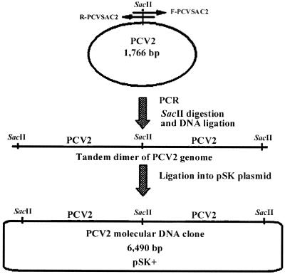
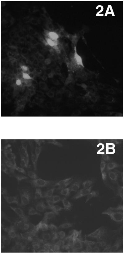
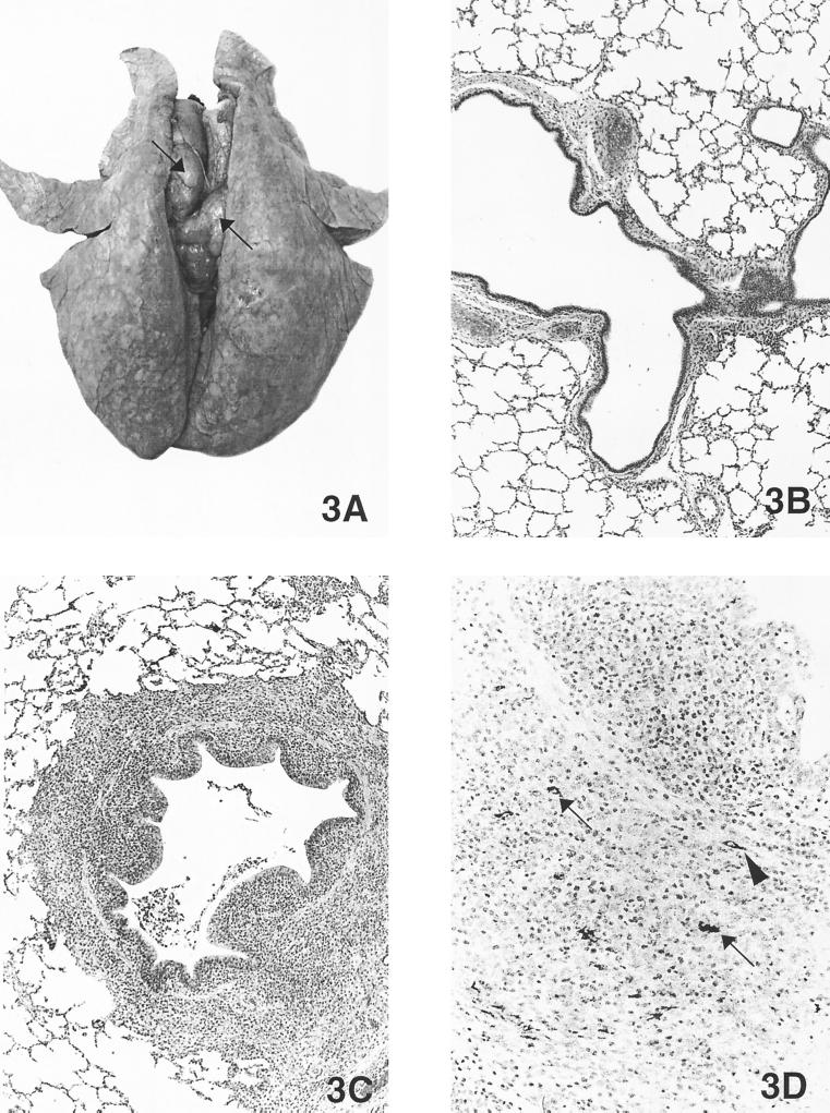
Infection of animals with a molecular viral clone is critical to study the genetic determinants of viral replication and virulence in the host. Type 2 porcine circovirus (PCV2) has been incriminated as the cause of postweaning multisystemic wasting syndrome (PMWS), an emerging disease in pigs. We report here for the first time the construction and use of an infectious molecular DNA clone of PCV2 to characterize the disease and pathologic lesions associated with PCV2 infection by direct in vivo transfection of pigs with the molecular clone. The PCV2 molecular clone was generated by ligating two copies of the complete PCV2 genome in tandem into the pBluescript SK (pSK) vector and was shown to be infectious in vitro when transfected into PK-15 cells. Forty specific-pathogen-free pigs at 4 weeks of age were randomly assigned to four groups of 10 each. Group 1 pigs served as uninoculated controls. Pigs in group 2 were each inoculated intranasally with about 1.9 x 10(5) 50% tissue culture infective doses of a homogeneous PCV2 live virus stock derived from the molecular clone. Pigs in group 3 were each injected intrahepatically with 200 microg of the cloned PCV2 plasmid DNA, and pigs in group 4 were each injected into the superficial iliac lymph nodes with 200 microg of the cloned PCV2 plasmid DNA. Animals injected with the cloned PCV2 plasmid DNA developed infection resembling that induced by intranasal inoculation with PCV2 live virus stock. Seroconversion to PCV2-specific antibody was detected in the majority of pigs from the three inoculated groups at 35 days postinoculation (DPI). Viremia, beginning at 14 DPI and lasting 2 to 4 weeks, was detected in the majority of the pigs from all three inoculated groups. There were no remarkable clinical signs of PMWS in control or any of the inoculated pigs. Gross lesions in pigs of the three inoculated groups were similar and were characterized by systemically enlarged, tan lymph nodes and lungs that failed to collapse. Histopathological lesions and PCV2-specific antigen were detected in numerous tissues and organs, including brain, lung, heart, kidney, tonsil, lymph nodes, spleen, ileum, and liver of infected pigs. This study more definitively characterizes the clinical course and pathologic lesions exclusively attributable to PCV2 infection. The data from this study indicate that the cloned PCV2 genomic DNA may replace infectious virus for future PCV2 pathogenesis and immunization studies. The data also suggest that PCV2, although essential for development of PMWS, may require other factors or agents to induce the full spectrum of clinical signs and lesions associated with advanced cases of PMWS.
Figures
Similar articles
-
Immunogenicity and pathogenicity of chimeric infectious DNA clones of pathogenic porcine circovirus type 2 (PCV2) and nonpathogenic PCV1 in weanling pigs.J Virol. 2003 Oct;77(20):11232-43. doi: 10.1128/jvi.77.20.11232-11243.2003. J Virol. 2003. PMID: 14512571 Free PMC article.
-
Experimental transmission of porcine circovirus type 2 (PCV2) in weaned pigs: a sequential study.J Comp Pathol. 2000 Nov;123(4):258-69. doi: 10.1053/jcpa.2000.0413. J Comp Pathol. 2000. PMID: 11041995
-
Effect of vaccination with selective bacterins on conventional pigs infected with type 2 porcine circovirus.Vet Pathol. 2003 Sep;40(5):521-9. doi: 10.1354/vp.40-5-521. Vet Pathol. 2003. PMID: 12949409 Clinical Trial.
-
Pathological findings associated with naturally acquired porcine circovirus type 2 associated disease.Vet Microbiol. 2004 Feb 4;98(2):137-49. doi: 10.1016/j.vetmic.2003.10.006. Vet Microbiol. 2004. PMID: 14741126 Review.
-
Porcine circovirus type 2 (PCV2): pathogenesis and interaction with the immune system.Annu Rev Anim Biosci. 2013 Jan;1:43-64. doi: 10.1146/annurev-animal-031412-103720. Epub 2013 Jan 3. Annu Rev Anim Biosci. 2013. PMID: 25387012 Review.
Cited by
-
Replacement of the replication factors of porcine circovirus (PCV) type 2 with those of PCV type 1 greatly enhances viral replication in vitro.J Virol. 2010 Sep;84(17):8986-9. doi: 10.1128/JVI.00522-10. Epub 2010 Jun 23. J Virol. 2010. PMID: 20573809 Free PMC article.
-
Immunogenicity and pathogenicity of chimeric infectious DNA clones of pathogenic porcine circovirus type 2 (PCV2) and nonpathogenic PCV1 in weanling pigs.J Virol. 2003 Oct;77(20):11232-43. doi: 10.1128/jvi.77.20.11232-11243.2003. J Virol. 2003. PMID: 14512571 Free PMC article.
-
Porcine circovirus type 2 and porcine circovirus-associated disease.J Vet Intern Med. 2009 Nov-Dec;23(6):1151-63. doi: 10.1111/j.1939-1676.2009.0389.x. Epub 2009 Sep 22. J Vet Intern Med. 2009. PMID: 19780932 Free PMC article. Review.
-
Effect of porcine circovirus type 2 (PCV2) vaccination of the dam on PCV2 replication in utero.Clin Vaccine Immunol. 2009 Jun;16(6):830-4. doi: 10.1128/CVI.00455-08. Epub 2009 Apr 8. Clin Vaccine Immunol. 2009. PMID: 19357312 Free PMC article.
-
Limited susceptibility of three different mouse (Mus musculus) lines to Porcine circovirus-2 infection and associated lesions.Can J Vet Res. 2009 Apr;73(2):81-6. Can J Vet Res. 2009. PMID: 19436587 Free PMC article.
References
-
- Allan, G. M., D. P. Mackie, J. McNair, B. M. Adair, and M. S. McNulty. 1994. Production, preliminary characterization and applications of monoclonal antibodies to porcine circovirus. Vet. Immunol. Immunopathol. 43:357–371. - PubMed
-
- Allan, G. M., F. McNeilly, J. P. Cassidy, G. A. Reilly, B. Adair, W. A. Ellis, and M. S. McNulty. 1995. Pathogenesis of porcine circovirus; experimental infections of colostrum deprived piglets and examination of pig fetal material. Vet. Microbiol. 44:49–64. - PubMed
-
- Allan, G. M., B. Meehan, D. Todd, S. Kennedy, F. McNeilly, J. Ellis, E. G. Clark, J. Harding, E. Espuna, A. Botner, and C. Charreyre. 1998. Novel porcine circoviruses from pigs with wasting disease syndromes. Vet. Rec. 142:467–468. - PubMed
-
- Allan, G. M., F. McNeilly, J. Ellis, S. Krakowka, B. Meeham, I. McNair, I. Walker, and S. Kennedy. 2000. Experimental infection of colostrums deprived piglets with porcine circovirus 2 (PCV2) and porcine reproductive and respiratory syndrome virus (PRRSV) potentiates PCV2 replication. Arch. Virol. 145:2421–2429. - PubMed
-
- Allan, G. M., F. McNeilly, S. Kennedy, B. Daft, E. G. Clarke, J. A. Ellis, D. M. Haines, B. M. Meehan, and B. M. Adair. 1998. Isolation of porcine circovirus-like viruses from pigs with a wasting disease in the USA and Europe. J. Vet. Diagn. Investig. 10:3–10. - PubMed
Publication types
MeSH terms
Substances
LinkOut - more resources
Full Text Sources
Other Literature Sources
Miscellaneous
