. 2002 Jan;76(2):569-81.
doi: 10.1128/jvi.76.2.569-581.2002.

A soluble factor(s) secreted from CD8(+) T lymphocytes inhibits human immunodeficiency virus type 1 replication through STAT1 activation

Affiliations

A soluble factor(s) secreted from CD8(+) T lymphocytes inhibits human immunodeficiency virus type 1 replication through STAT1 activation

Theresa Li-Yun Chang et al. J Virol. 2002 Jan.

Abstract

CD8(+) T lymphocytes can suppress human immunodeficiency virus type 1 (HIV-1) replication by secreting a soluble factor(s) known as CD8(+) T-lymphocyte antiviral factor (CAF). One site of CAF action is inhibition of HIV-1 RNA transcription, particularly at the step of long terminal repeat (LTR)-driven gene expression. However, the mechanism by which CAF inhibits LTR activation is not understood. Here, we show that conditioned media from several herpesvirus saimari-transformed CD8(+) T lymphocytes inhibit, in a time- and dose-dependent manner, the replication of HIV-1 pseudotype viruses that express the envelope glycoproteins of vesicular stomatitis virus (HIV-1(VSV)). The same conditioned media also inhibit phorbol myristate acetate-induced activation of the HIV-1 LTR and activate the signal transducer and activator of transcription 1 (STAT1) protein. We have obtained direct evidence that STAT1 is necessary for CAF-mediated inhibition of LTR activation and HIV-1 replication. Thus, the inhibitory effect of CAF on HIV-1(VSV) replication was abolished in STAT1-deficient cells. Moreover, CAF inhibition of LTR activation was diminished both in STAT1-deficient cells and in cells expressing a STAT1 dominant negative mutant but was restored when STAT1 was reintroduced into the STAT1-deficient cells. We also observed that CAF induced the expression of interferon regulatory factor 1 (IRF-1), and that IRF-1 gene induction was STAT-1 dependent. Taken together, our results suggest that CAF activates STAT1, leading to IRF-1 induction and inhibition of gene expression regulated by the HIV-1 LTR. This study therefore helps clarify one molecular mechanism of host defense against HIV-1.


Figures

FIG. 1.
FIG. 1.
Time- and dose-dependent inhibition of HIV-1 infection by CAF. (A) Primary macrophages were infected with HIV-1 BaL and then treated with a 10% concentration of CAF from the indicated CD8+-T-cell lines or with no CAF (complete medium). The extracellular HIV-1 p24 antigen concentration was measured on day 10 postinfection. (B) HeLa cells were treated with a 50% concentration of CAF from the K#1 50K line for the indicated periods before infection with luciferase-expressing HIV-1VSV. (C) HeLa cells were treated with different dilutions of the K#1 50K conditioned medium for 16 h before HIV-1VSV infection. In both experiments, luciferase activities were measured at 48 h postinfection. In panels B and C, the results (mean ± standard error) from triplicate determinations in a single experiment are presented as histograms. Similar results were obtained in another independent experiment. The values represent luciferase activity compared to that derived from cells not treated with CAF, which was defined as 100%. This corresponded to absolute r.l.u. values of 810 ± 80. In panel B, the difference between cells treated with CAF for 16 h and nontreated cells is significant (P = 0.025). In panel C, the difference between cells treated with 50% CAF and nontreated cells is significant (P = 0.023).
FIG. 2.
FIG. 2.
Effects of conditioned medium from HVS-transformed CD8+ T cells on HIV-1 LTR activation. (A) (Left panel) 1G5 cells, which stably express the HIV-LTR-Luc construct, were treated for 16 h with or without a 50% concentration of CAF (K#1 50K line), before addition of PMA for 8 h. The P value for the difference between the values obtained from cells treated with 50% CAF and the control cells is 0.012. (Right panel) Cells were treated for 8 h with or without CAF, added simultaneously with PMA. (B) The experiment was as described for panel A, except that HeLa cells were transiently transfected with the HIV-LTR-Luc plasmid for 16 h before treatment with CAF and/or PMA. In both panels A and B, the results (mean ± standard error) from triplicate determinations in a single experiment are presented as histograms. Similar results were obtained in two other independent experiments. The values represent luciferase activity compared to that derived from cells not treated with CAF, which was defined as 100%. This corresponded to absolute r.l.u. values of 243 ± 12 for 1G5 cells and 2,319 ± 41 for HeLa cells. The background values for 1G5 and HeLa cells are 1.5 ± 0.4. Luciferase activities were normalized for cellular protein content (1G5 cells) or β-galactosidase activity (HeLa cells). (C) HeLa cells were transiently transfected with HIV-LTR-Luc and then treated with conditioned media from different HVS-transformed CD8+-T-cell clones for a further 16 h. Luciferase activity was measured after PMA stimulation for 8 h.
FIG. 3.
FIG. 3.
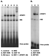
STAT1 activation is induced by CAF from different HVS-transformed CD8+-T-cell lines. 1G5 cells were treated for 15 min with a 50% concentration of conditioned medium from the indicated lines. Whole-cell extracts were prepared for EMSA using a 32P-labeled m67 GAS probe. The presence of STAT1 in the complexes was confirmed by adding anti-STAT1 antibody in a supershift experiment (panel A, lane 6).
FIG. 4.
FIG. 4.
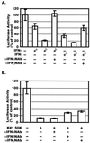
Effect of CAF on PMA-induced HIV-1 LTR activation in the presence of neutralizing antibodies against IFN-α, IFN-β, or IFN-γ. (A) HeLa cells were transiently transfected with the HIV-LTR-Luc plasmid and then treated with 100 U (+a) or 1,000 U (+b) of IFN-α per ml or 20 ng (+c) or 200 ng (+d) of IFN-γ per ml for 16 h. Neutralizing antibodies, when present (dilution of stocks,1:250), were incubated with the IFN stocks for 30 min at 37°C before addition of the mixture to the cells. Luciferase activities were measured after stimulation of the cells with PMA for 8 h. (B) The experiment was performed as described for panel A except that CAF (50% conditioned medium from line K#1 50K) replaced the IFNs. The results (mean ± standard error) are presented as histograms. They represent luciferase activity compared to the value derived from cells not treated with CAF or IFN, which was defined as 100%. This corresponded to absolute r.l.u. values of 2,140 ± 374.
FIG. 5.
FIG. 5.
Effects of CAF on HIV-1VSV replication and STAT1 activation in U3A and 2fTGH cells. (A) 2fTGH (STAT1-expressing) and U3A (STAT1-null) cells were incubated with (solid bar) or without (open bar) a 50% concentration of CAF (K#1 50K clone) for 16 h before infection with HIV-1VSV. The results (mean ± standard error) from triplicate determinations in a single experiment are presented as histograms. Similar results were obtained in two other independent experiments. The values represent luciferase activity compared to that derived from cells not treated with CAF, which was defined as 100%. This corresponded to absolute r.l.u. values of 11,391 ± 393 for 2fTGH and 18,600 ± 500 for U3A. The average background r.l.u. value was 1.5. (B) U3A and 2fTGH cells were treated with 50% CAF (K#1 50K clone) for 15 min before whole-cell extracts were prepared for EMSA using a 32P-labeled m67 GAS probe.
FIG. 5.
FIG. 5.
Effects of CAF on HIV-1VSV replication and STAT1 activation in U3A and 2fTGH cells. (A) 2fTGH (STAT1-expressing) and U3A (STAT1-null) cells were incubated with (solid bar) or without (open bar) a 50% concentration of CAF (K#1 50K clone) for 16 h before infection with HIV-1VSV. The results (mean ± standard error) from triplicate determinations in a single experiment are presented as histograms. Similar results were obtained in two other independent experiments. The values represent luciferase activity compared to that derived from cells not treated with CAF, which was defined as 100%. This corresponded to absolute r.l.u. values of 11,391 ± 393 for 2fTGH and 18,600 ± 500 for U3A. The average background r.l.u. value was 1.5. (B) U3A and 2fTGH cells were treated with 50% CAF (K#1 50K clone) for 15 min before whole-cell extracts were prepared for EMSA using a 32P-labeled m67 GAS probe.
FIG. 6.
FIG. 6.
Effects of CAF on HIV-1 LTR activation in HeLa and U3A cells expressing a dominant negative STAT1 protein. (A) HeLa cells were transiently transfected with a plasmid expressing HIV-LTR-Luc and one expressing either no STAT1 (vector control), wild-type STAT1, or the STAT1 Y701F dominant negative mutant (STAT1 DN), as indicated. The transfected cells were then treated with CAF (50% concentration; K#1 50K line) for 16 h before PMA stimulation for 8 h and measurement of luciferase activity. (B) A similar experiment was performed using U3A (STAT1-null) cells. The results (mean ± standard error) from triplicate determinations in a single experiment are presented as histograms. Similar results were obtained in another independent experiment. The values represent luciferase activity compared to that derived from cells not treated with CAF, which was defined as 100%. This corresponded to absolute r.l.u. values of 1,850 ± 50 for HeLa cells and 4,650 ± 13 for U3A cells.
FIG. 7.
FIG. 7.
IRF-1 was induced and bound to DBF sites within the HIV-1 LTR as a response to CAF. Different cell lines (A, 1G5; B, 2fTGH, U3A, and HeLa) were treated with CAF (50% concentration; K#1 50K line) for 16 h. Whole-cell extracts were prepared for EMSA using a 32P-labeled HIV-1 DBF probe. The composition of protein complexes was further assessed by adding anti-IRF-1, anti-IRF-2 or anti-STAT1 antibodies in a supershift experiment, as indicated. (C) HeLa cells were treated with conditioned media (50% concentration) from lines KP1#3 and K#1 50K, as indicated. Anti-IRF-1 or anti-IRF-2 antibodies were added in a supershift experiment, as indicated. X denotes a complex of unknown composition that is induced by all conditioned media, irrespective of the presence of CAF activity (see text). (D) Whole-cell extracts from 1G5 cells treated with CAF (50% concentration; K1 50K line) were analyzed by EMSA using a 32P-labeled ISG15 probe. Anti-IRF-1 or anti-IRF-2 antibodies were added in a supershift experiment, as indicated.

Similar articles

Cited by

References

    1. Bailer, R. T., A. Holloway, J. Sun, J. B. Margolick, M. Martin, J. Kostman, and L. J. Montaner. 1999. IL-13 and IFN-gamma secretion by activated T cells in HIV-1 infection associated with viral suppression and a lack of disease progression. J. Immunol. 162:7534–7542. - PubMed
    1. Barker, E., K. N. Bossart, and J. A. Levy. 1998. Primary CD8+ cells from HIV-infected individuals can suppress productive infection of macrophages independent of beta-chemokines. Proc. Natl. Acad. Sci. USA 95:1725–1729. - PMC - PubMed
    1. Benkirane, M., R. F. Chun, H. Xiao, V. V. Ogryzko, B. H. Howard, Y. Nakatani, and K. T. Jeang. 1998. Activation of integrated provirus requires histone acetyltransferase: p300 and P/CAF are coactivators for HIV-1 Tat. J. Biol. Chem. 273:24898–24905. - PubMed
    1. Blanco, J. C., C. Contursi, C. A. Salkowski, D. L. DeWitt, K. Ozato, and S. N. Vogel. 2000. Interferon regulatory factor (IRF)-1 and IRF-2 regulate interferon gamma-dependent cyclooxygenase 2 expression. J. Exp. Med. 191:2131–2144. - PMC - PubMed
    1. Bovolenta, C., L. Camorali, A. L. Lorini, S. Ghezzi, E. Vicenzi, A. Lazzarin, and G. Poli. 1999. Constitutive activation of STATs upon in vivo human immunodeficiency virus infection. Blood 94:4202–4209. - PubMed

Publication types

MeSH terms