Comparative Study
. 2002 Jan;76(2):609-18.
doi: 10.1128/jvi.76.2.609-618.2002.

Sequential partially overlapping gene arrangement in the tricistronic S1 genome segments of avian reovirus and Nelson Bay reovirus: implications for translation initiation

Affiliations
Comparative Study

Sequential partially overlapping gene arrangement in the tricistronic S1 genome segments of avian reovirus and Nelson Bay reovirus: implications for translation initiation

Maya Shmulevitz et al. J Virol. 2002 Jan.

Abstract

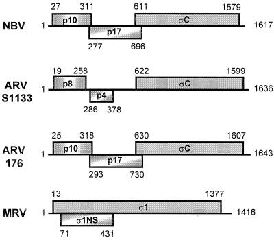
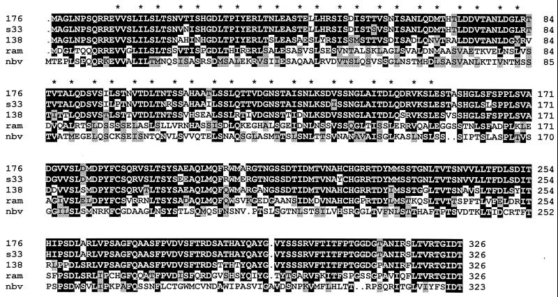
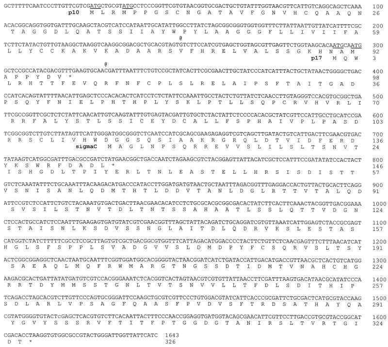
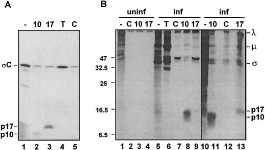
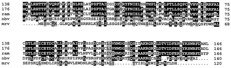
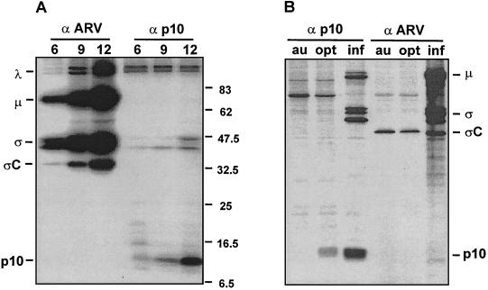
Previous studies of the avian reovirus strain S1133 (ARV-S1133) S1 genome segment revealed that the open reading frame (ORF) encoding the final sigmaC viral cell attachment protein initiates over 600 nucleotides distal from the 5' end of the S1 mRNA and is preceded by two predicted small nonoverlapping ORFs. To more clearly define the translational properties of this unusual polycistronic RNA, we pursued a comparative analysis of the S1 genome segment of the related Nelson Bay reovirus (NBV). Sequence analysis indicated that the 3'-proximal ORF present on the NBV S1 genome segment also encodes a final sigmaC homolog, as evidenced by the presence of an extended N-terminal heptad repeat characteristic of the coiled-coil region common to the cell attachment proteins of reoviruses. Most importantly, the NBV S1 genome segment contains two conserved ORFs upstream of the final sigmaC coding region that are extended relative to the predicted ORFs of ARV-S1133 and are arranged in a sequential, partially overlapping fashion. Sequence analysis of the S1 genome segments of two additional strains of ARV indicated a similar overlapping tricistronic gene arrangement as predicted for the NBV S1 genome segment. Expression analysis of the ARV S1 genome segment indicated that all three ORFs are functional in vitro and in virus-infected cells. In addition to the previously described p10 and final sigmaC gene products, the S1 genome segment encodes from the central ORF a 17-kDa basic protein (p17) of no known function. Optimizing the translation start site of the ARV p10 ORF lead to an approximately 15-fold increase in p10 expression with little or no effect on translation of the downstream final sigmaC ORF. These results suggest that translation initiation complexes can bypass over 600 nucleotides and two functional overlapping upstream ORFs in order to access the distal final sigmaC start site.


Figures

FIG. 1.
FIG. 1.
Gene arrangement of the polycistronic S1 genome segments of orthoreoviruses. The arrangement of predicted ORFs present in the ARV-176, ARV-S1133, NBV, and MRV S1 genome segments is depicted. The ORFs are shown as shaded boxes above and below a line representing the mRNA. The ORFs are identified by the name or approximate molecular mass (in kilodaltons) of the predicted gene product. Numbers indicate the first and last nucleotide numbers of each ORF (including the initiation codon, excluding the termination codon). The ARV-138 gene arrangement is identical to that depicted for ARV-176. The ARV-S1133 gene arrangement is based on the previously published sequence (46), while the MRV gene arrangement depicted is based on the published sequence of strain Dearing (12).
FIG. 2.
FIG. 2.
Multiple sequence alignment of the reovirus cell attachment proteins. The predicted amino acid sequences of the two ARV ςC proteins determined in this study (ARV-176 [176]) and ARV-138 [138]) and of the gene product of the 3′-proximal ORF of NBV (nbv) were aligned with the predicted sequences of the ςC cell attachment proteins of two previously sequenced ARV isolates (ARV-S1133 [s33] and ARV-Ram1 [ram]) using PILEUP and shaded using BOXSHADE. The asterisks denote the locations of the apolar residues present in the conserved heptad repeat structure. Identical residues present in three or more of the aligned sequences are indicated by black background shading, and grey shading denotes conservation of similar residues. Dots indicate insertions.
FIG. 3.
FIG. 3.
ARV-176 S1 genome segment cDNA sequence and predicted amino acid sequences of the encoded gene products. The complete cDNA plus-strand sequence of the ARV-176 S1 genome segment is presented, along with the amino acid sequences of the encoded p10, p17, and ςC proteins. Asterisks indicate the locations of the termination codons of the three ORFs. Potential start codons lying upstream of the ςC translation start site are underlined. The two @ symbols above the cDNA sequence denote the locations of single-base insertions, relative to the published ARV-S1133 sequence, that change the reading frame of the p10 and p17 ORFs (see text).
FIG. 4.
FIG. 4.
ARV S1 mRNA is functionally tricistronic in vitro and in vivo. (A) The full-length ARV-176 S1 cDNA clone was used for the production of capped mRNA by in vitro transcription. The transcripts were translated in rabbit reticulocyte lysates, and the [3H]leucine-labeled translation mixtures were fractionated by SDS-PAGE (15% acrylamide) either without immunoprecipitation (−) or after precipitation with the monospecific anti-p10 (10) or anti-p17 (17) serum, with antiserum raised against total virus structural proteins to detect ςC (T), or with control preimmune serum (C). Radiolabeled polypeptides were detected by fluorography. The locations of the ςC, p17, and p10 gene products are indicated on the left. (B) Uninfected (uninf., lanes 1 to 4) or ARV-infected (inf., lanes 5 to 13) QM5 cells were pulse-labeled with [3H]leucine for 1 h at 16 h postinfection. Cell lysates were fractionated by SDS-PAGE (15% acrylamide) either without immunoprecipitation (−) or after precipitation with the same antisera described for panel A. The locations of molecular size markers are indicated on the left (in kilodaltons), and the locations of the p10, p17, and major λ, μ, and ς virus structural proteins are indicated on the right.
FIG. 5.
FIG. 5.
Multiple sequence alignment of the p17 and ς1NS proteins. The predicted amino acid sequences of the two ARV p17 proteins (ARV-176 [176]) and ARV-138 [138]) and of the NBV p17 protein (nbv) determined in this study were aligned with the predicted sequences of the p17 protein of a previously sequenced ARV isolate (ARV-Ram1 [ram]) and with the ς1NS protein of mammalian reovirus strain Dearing (mrv) using PILEUP and shaded using BOXSHADE. Identical residues present in three or more of the aligned sequences are indicated by black background shading, and grey shading denotes conservation of similar residues. Dots indicate insertions.
FIG. 6.
FIG. 6.
Translation analysis of the ARV S1 genome segment. (A) ARV-infected cells were pulse-labeled for 1 h using [35S]methionine at 6, 9, or 12 h postinfection. Infected cell lysates were immunoprecipitated using polyclonal antiserum specific for either ARV structural proteins, including ςC (α ARV) or the nonstructural p10 protein (α p10). Precipitates were fractionated by SDS-PAGE (15% acrylamide), and the labeled products were detected by autoradiography. The locations of molecular mass standards are indicated on the right (in kilodaltons). The locations of the major species of λ-, μ-, and ς-class ARV proteins are indicated on the left, along with the locations of the ςC and p10 gene products encoded by the polycistronic S1 genome segment. (B) QM5 cell monolayers were either infected with ARV-176 (inf) or transfected with plasmids expressing the S1 genome segment of ARV-176 containing either an authentic (au) or optimized (opt) p10 translation start site. Monolayers were labeled with [35S]methionine at 16 h postinfection or 36 h posttransfection, and the radiolabeled cell lysates were immunoprecipitated as described for panel A. Precipitates were fractionated by SDS-PAGE (15% acrylamide), and the labeled products were detected by autoradiography. The locations of the major species of μ- and ς-class ARV proteins are indicated on the right, along with the locations of the ςC and p10 gene products. Higher-molecular-weight species in lane 3 represent nonspecific trapping of radiolabeled virus particles by the precipitation reaction.

Similar articles

Cited by

References

    1. Ahmadian, G., J. S. Randhawa, and A. J. Easton. 2000. Expression of the ORF-2 protein of the human respiratory syncytial virus M2 gene is initiated by a ribosomal termination-dependent reinitiation mechanism. EMBO J. 19:2681–2689. - PMC - PubMed
    1. Ahne, W., I. Thomsen, and J. Winton. 1987. Isolation of a reovirus from the snake Python regius. Arch. Virol. 94:135–139. - PubMed
    1. Antin, P. B., and C. P. Ordahl. 1991. Isolation and characterization of an avian myogenic cell line. Dev. Biol. 143:111–121. - PubMed
    1. Chappell, J. D., V. L. Gunn, J. D. Wetzel, G. S. Baer, and T. S. Dermody. 1997. Mutations in type 3 reovirus that determine binding to sialic acid are contained in the fibrous tail domain of viral attachment protein sigma1. J. Virol. 71:1834–1841. - PMC - PubMed
    1. Chappell, J. D., J. L. Duong, B. W. Wright, and T. S. Dermody. 2000. Identification of carbohydrate-binding domains in the attachment proteins of type 1 and type 3 reoviruses. J. Virol. 74:8472–8479. - PMC - PubMed

Publication types

MeSH terms