

The simian immunodeficiency virus deltaNef vaccine, after application to the tonsils of Rhesus macaques, replicates primarily within CD4(+) T cells and elicits a local perforin-positive CD8(+) T-cell response
- PMID: 11752159
- PMCID: PMC136843
- DOI: 10.1128/jvi.76.2.688-696.2002
The simian immunodeficiency virus deltaNef vaccine, after application to the tonsils of Rhesus macaques, replicates primarily within CD4(+) T cells and elicits a local perforin-positive CD8(+) T-cell response
Abstract
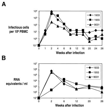
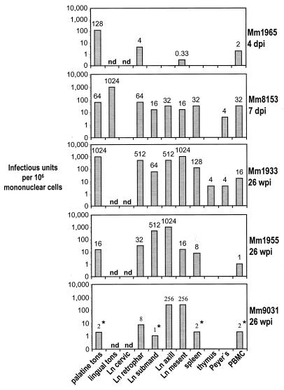
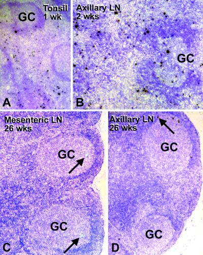
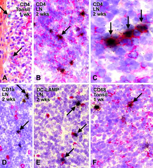
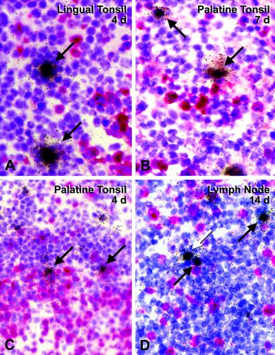
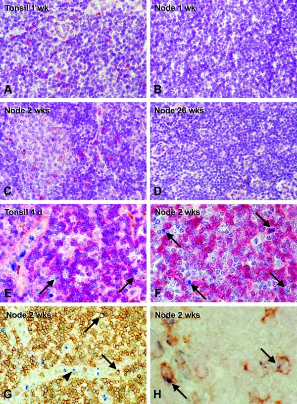
Deletion of the nef gene from simian immunodeficiency virus (SIV) strain SIVmac239 yields a virus that undergoes attenuated growth in rhesus macaques and offers substantial protection against a subsequent challenge with some SIV wild-type viruses. We used a recently described model to identify sites in which the SIVDeltanef vaccine strain replicates and elicits immunity in vivo. A high dose of SIVDeltanef was applied to the palatine and lingual tonsils, where it replicated vigorously in this portal of entry at 7 days. Within 2 weeks, the virus had spread and was replicating actively in axillary lymph nodes, primarily in extrafollicular T-cell-rich regions but also in germinal centers. At this time, large numbers of perforin-positive cells, both CD8(+) T cells and CD3-negative presumptive natural killer cells, were found in the tonsil and axillary lymph nodes. The number of infected cells and perforin-positive cells then fell. When autopsy studies were carried out at 26 weeks, only 1 to 3 cells hybridized for viral RNA per section of lymphoid tissue. Nevertheless, infected cells were detected chronically in most lymphoid organs, where the titers of infectious virus could exceed by a log or more the titers in blood. Immunocytochemical labeling at the early active stages of infection showed that cells expressing SIVDeltanef RNA were CD4(+) T lymphocytes. A majority of infected cells were not in the active cell cycle, since 60 to 70% of the RNA-positive cells in tissue sections lacked the Ki-67 cell cycle antigen, and both Ki-67-positive and -negative cells had similar grain counts for viral RNA. Macrophages and dendritic cells, identified with a panel of monoclonal antibodies to these cells, were rarely infected. We conclude that the attenuated growth and protection observed with the SIVDeltanef vaccine strain does not require that the virus shift its characteristic site of replication, the CD4(+) T lymphocyte. In fact, this immunodeficiency virus can replicate actively in CD4(+) T cells prior to being contained by the host, at least in part by a strong killer cell response that is generated acutely in the infected lymph nodes.
Figures
References
-
- Amara, R. R., F. Villinger, J. D. Altman, S. L. Lydy, S. P. O’Neil, S. I. Staprans, D. C. Montefiori, Y. Xu, J. G. Herndon, L. S. Wyatt, M. A. Candido, N. L. Kozyr, P. L. Earl, J. M. Smith, H.-L. Ma, B. D. Grimm, M. L. Hulsey, J. Miller, H. M. McClure, J. M. McNicholl, B. Moss, and H. L. Robinson. 2001. Control of mucosal challenge and prevention of AIDS by a multiprotein DNA/MVA vaccine. Science 292:69–74. - PubMed
-
- Baba, T. W., A. M. Trichel, L. An, V. Liska, L. N. Martin, M. Murphey-Corb, and R. M. Ruprecht. 1996. Infection and AIDS in adult macaques after nontraumatic oral exposure to cell-free SIV. Science 272:1486–1489. - PubMed
-
- Barouch, D. H., S. Santra, J. E. Schmitz, M. J. Kuroda, T. M. Fu, W. Wagner, M. Bilska, A. Craiu, X. X. Zheng, G. R. Krivulka, K. Beaudry, M. A. Lifton, C. E. Nickerson, W. L. Trigona, K. Punt, D. C. Freed, L. Guan, S. Dubey, D. Casimiro, A. Simon, M. E. Davies, M. Chastain, T. B. Strom, R. S. Gelman, D. C. Montefiori, and M. G. Lewis. 2000. Control of viremia and prevention of clinical AIDS in rhesus monkeys by cytokine-augmented DNA vaccination. Science 290:486–492. - PubMed
-
- Chakrabarti, L., V. Baptiste, E. Khatissian, M.-C. Cumont, A.-M. Aubertin, L. Montagnier, and B. Hurtel. 1995. Limited viral spread and rapid immune response in lymph nodes of macaques inoculated with attenuated simian immunodeficiency virus. Virology 213:535–548. - PubMed
Publication types
MeSH terms
Substances
Grants and funding
LinkOut - more resources
Full Text Sources
Research Materials
