. 2002 Jan;76(2):841-50.
doi: 10.1128/jvi.76.2.841-850.2002.

Herpes simplex virus type 1 immediate-early protein ICP0 and is isolated RING finger domain act as ubiquitin E3 ligases in vitro

Affiliations

Herpes simplex virus type 1 immediate-early protein ICP0 and is isolated RING finger domain act as ubiquitin E3 ligases in vitro

Chris Boutell et al. J Virol. 2002 Jan.

Abstract

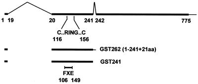
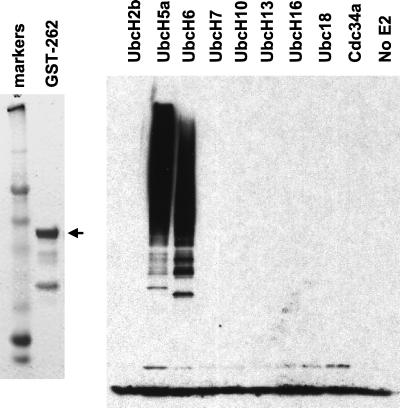
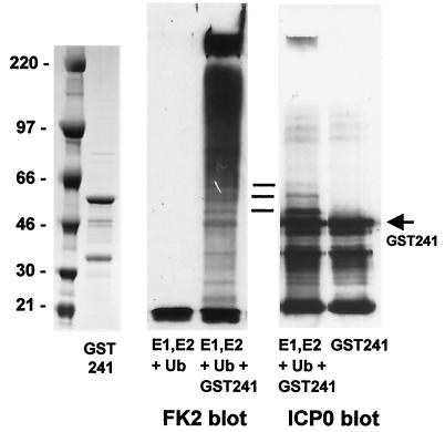
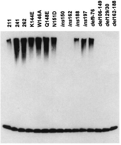
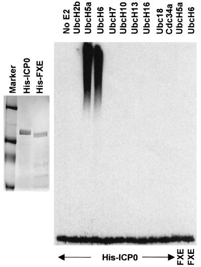
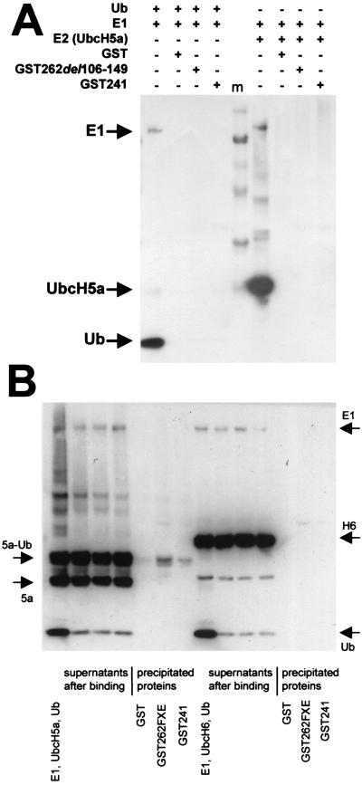
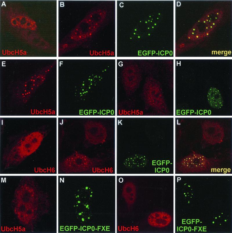
Proteasome-dependent degradation of ubiquitinated proteins plays a key role in many important cellular processes. Ubiquitination requires the E1 ubiquitin activating enzyme, an E2 ubiquitin conjugating enzyme, and frequently a substrate-specific ubiquitin protein ligase (E3). One class of E3 ubiquitin ligases has been shown to contain a common zinc-binding RING finger motif. We have previously shown that herpes simplex virus type 1 ICP0, itself a RING finger protein, induces the proteasome-dependent degradation of several cellular proteins and induces the accumulation of colocalizing conjugated ubiquitin in vivo. We now report that both full-length ICP0 and its isolated RING finger domain induce the accumulation of polyubiquitin chains in vitro in the presence of E1 and the E2 enzymes UbcH5a and UbcH6. Mutations within the RING finger region that abolish the in vitro ubiquitination activity also cause severe reductions in ICP0 activity in other assays. We conclude that ICP0 has the potential to act as an E3 ubiquitin ligase during viral infection and to target specific cellular proteins for destruction by the 26S proteasome.


Figures

FIG. 1.
FIG. 1.
Map of the ICP0 transcript and coding region. The upper line shows the ICP0 coding region (thick line), the introns (thin inverted v-lines), and the 3′ noncoding region (thin line). The numbers refer to the codon positions at the intron junctions and the last coding triplet. The position of the RING finger in exon 2 is indicated with the locations of the first and last zinc-coordinating cysteine residues. Below the map are the ICP0 residues expressed as a GST fusion protein. GST-262 includes residues 1 to 241 encoded in exons 1 and 2 (thick line) plus 21 residues derived from intron 2 sequences. The ICP0 portion of the GST fusion protein is equivalent to ICP0-262 (ICP0R). GST-241 includes only ICP0 residues 1 to 241. The position of the RING finger deletion mutation in clones derived from mutant FXE is indicated.
FIG. 2.
FIG. 2.
Typical expression and purification of a GST-ICP0 RING finger domain fusion protein and its activity in an in vitro ubiquitin conjugation assay. GST-262 was expressed in bacteria and purified using glutathione-agarose beads as described in Materials and Methods. The left panel shows a Coomassie-stained gel of the fusion protein that migrates at approximately 60 kDa (arrow). The right panel shows the results of an in vitro ubiquitin conjugation assay, probed using antiubiquitin antibody P4D1, as described in the text. A panel of nine different E2 enzymes were screened for activity in the presence of GST-262. The related proteins UbcH5a and UbcH6 were stimulated by GST-262, but the others were not.
FIG. 3.
FIG. 3.
Activity and autoubiquitination of a GST fusion protein including the first 241 residues of ICP0. The left panel shows a Coomassie-stained gel of purified GST-241. The middle panel shows the in vitro ubiquitination activity of the purified GST-241 protein (right) next to a control sample without added GST-241 (left), as detected with antibody FK2 that detects conjugated ubiquitin. The right panel shows a portion of the same filter reprobed to detect GST-241 after incubation with E1, UbcH5a, and ubiquitin (left), next to a control untreated sample (right). The horizontal lines indicate modified forms of GST-241 that comigrate with minor conjugated ubiquitin bands and that appear only after incubation in the in vitro conjugation reaction mixture.
FIG. 4.
FIG. 4.
Comparison of the effects of mutations in the first and second exons of ICP0 in the in vitro conjugation reaction. GST fusion proteins based on either GST-262 or GST-241 were purified after bacterial expression. The locations and phenotypes of the mutations are shown in Table 1. All the proteins had similar expression, solubility, and stability properties (data not shown). The products of the in vitro ubiquitin conjugation reaction using E1, ubiquitin, UbcH5a, and added GST fusion protein were detected by probing a Western blot with the antiubiquitin antibody P4D1.
FIG. 5.
FIG. 5.
Full-length ICP0 stimulates the activity of UbcH5a and UbcH6 in vitro. Wild-type and RING finger mutant full-length ICP0 proteins with N-terminal His-tags were expressed in insect cells using a recombinant baculovirus and purified by nickel affinity chromatography. The left panel shows typical samples of purified His-ICP0 and His-FXE, stained by Coomassie blue. The right panel shows the results of a screen for stimulation of the activity of a panel of E2 ubiquitin conjugation enzymes, probed for high-molecular-weight ubiquitin conjugates using antibody P4D1. Like the GST fusion constructs containing ICP0 RING domains, His-ICP0 stimulates the activity of UbcH5a and UbcH6. The rightmost two tracks show a similar assay using purified RING finger deletion mutant ICP0 (FXE) with UbcH5a and UbcH6.
FIG. 6.
FIG. 6.
ICP0 does not form a stable complex in vitro with E1, ubiquitin, UbcH5a, UbcH6, or the UbcH5a-ubiquitin thiol ester. (A) Purified His-E1 and His-ubiquitin (lefthand set of tracks) or His-E1 and His-UbcH5a (righthand set of tracks) were preincubated at 37°C for 15 min in E3 ligase reaction buffer and then precleared by incubation with glutathione-agarose beads charged with GST for 10 min on ice. Glutathione-agarose beads charged with GST, GST-241, and the RING finger mutant GST-262(del106-149) (FXE) were prepared and incubated on ice for 20 min with aliquots of the respective mixtures. The beads were washed five times with 50 mM Tris-HCl (pH 7.5), 100 mM NaCl, 5% glycerol, and 0.05% NP-40 and then pelleted, and coprecipitating proteins were released by boiling in SDS gel loading buffer without reducing agent, so as to preserve any ubiquitin-thiol ester linkages. The samples were analyzed by SDS-PAGE followed by Western blotting with an antipolyhistidine antibody. Each set of four tracks shows, from left to right, a sample of the target reaction mixture and proteins in the pellets of GST, GST-262FXE, and GST-241 beads, respectively. The sets are separated by molecular weight markers (m), and the ingredients in each incubation mix are indicated above the gel lanes. (B) The lefthand set of seven tracks shows the results from a similar experiment in which the UbcH5a-ubiquitin thiol ester intermediate was prepared by incubating E1, ubiquitin, and UbcH5a in E3 ligase buffer at 37°C for 15 min. All subsequent operations were performed on ice to preserve the thiol ester linkage. After preclearing the initial mixture as above, aliquots were incubated with beads charged with GST, GST-241, and the RING finger deletion derivative and then the samples were washed and analyzed as above. From left to right the tracks show the initial reaction mixture, the supernatants, and the pellets after binding. Equivalent proportions of the total bead and supernatant fractions were loaded onto the gel to illustrate the true relative amounts of bound and unbound protein. Although some UbcH5a-Ub was present in the pellets, this was minor compared to the unbound fraction and was also present in the negative control. The righthand part of the panel shows a similar experiment with UbcH6. In this example, the formation of the UbcH6-ubiquitin thiol ester linkage was considerably less efficient than that with UbcH5a.
FIG. 7.
FIG. 7.
ICP0 sequesters UbcH5a and UbcH6 in transfected cells in a RING finger-dependent manner. HEp-2 cells were transfected with plasmids expressing tagged UbcH5a or UbcH6 and EGFP-linked or native ICP0 and ICP0 RING finger mutant derivatives. The E2 enzymes were detected by the UL30 tag (r113) (UbcH5a) or the FLAG-tag (UbcH6) and the appropriate Cy5-conjugated secondary antibodies. (A) The Cy5-labeled UbcH5a single transfection control. (B to D) The single and merged channels for a cell coexpressing UbcH5a and EGFP-ICP0. (E, F, G, and H) The separated channels of further examples of cotransfected wild-type EGFP-ICP0-expressing cells. (I) A typical cell expressing UbcH6 alone. (J to L) The separated and merged channels of an image of a singly UbcH6-transfected cell (top) and one doubly transfected with EGFP-ICP0. (M and N) A cell expressing UbcH5a and ICP0 RING finger mutant FXE (del 106–149) linked to EGFP. (O and P) A similar experiment using UbcH6.

Similar articles

Cited by

References

    1. Chelbi-Alix, M. K., and H. de The. 1999. Herpes virus induced proteasome-dependent degradation of the nuclear bodies-associated PML and Sp100 proteins. Oncogene 18:935–941. - PubMed
    1. Everett, R. D. 1987. A detailed mutational analysis of Vmw110, a trans-acting transcriptional activator encoded by herpes simplex virus type 1. EMBO J. 6:2069–2076. - PMC - PubMed
    1. Everett, R. D. 1988. Analysis of the functional domains of herpes simplex virus type 1 immediate-early polypeptide Vmw110. J. Mol. Biol. 202:87–96. - PubMed
    1. Everett, R. D. 2000. ICP0 induces the accumulation of colocalizing conjugated ubiquitin. J. Virol. 74:9994–10005. - PMC - PubMed
    1. Everett, R. D. 2000. ICP0, a regulator of herpes simplex virus during lytic and latent infection. Bioessays 22:761–770. - PubMed

Publication types

MeSH terms