. 2002 Jan;76(2):875-84.
doi: 10.1128/jvi.76.2.875-884.2002.

Dominance of CD8 responses specific for epitopes bound by a single major histocompatibility complex class I molecule during the acute phase of viral infection

Affiliations

Dominance of CD8 responses specific for epitopes bound by a single major histocompatibility complex class I molecule during the acute phase of viral infection

Bianca R Mothé et al. J Virol. 2002 Jan.

Abstract

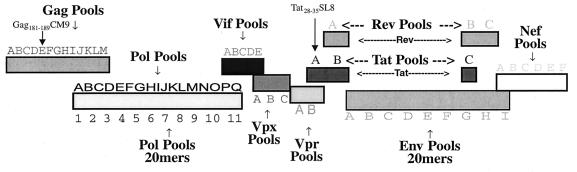
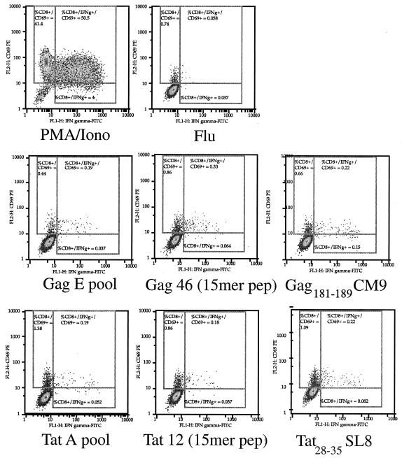
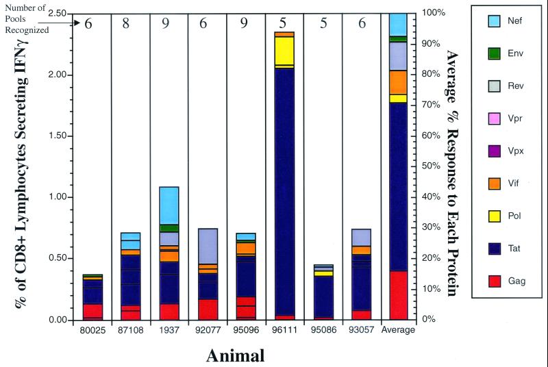
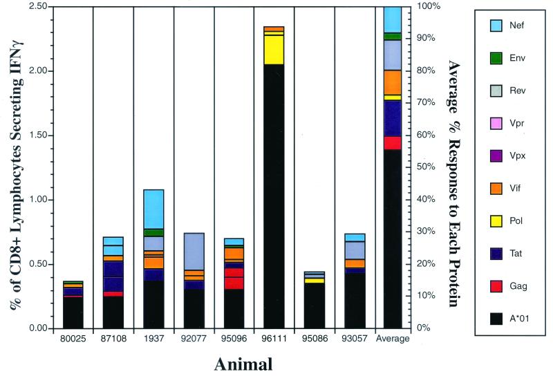
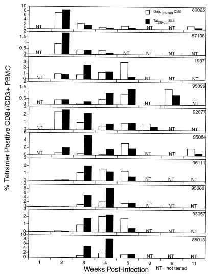
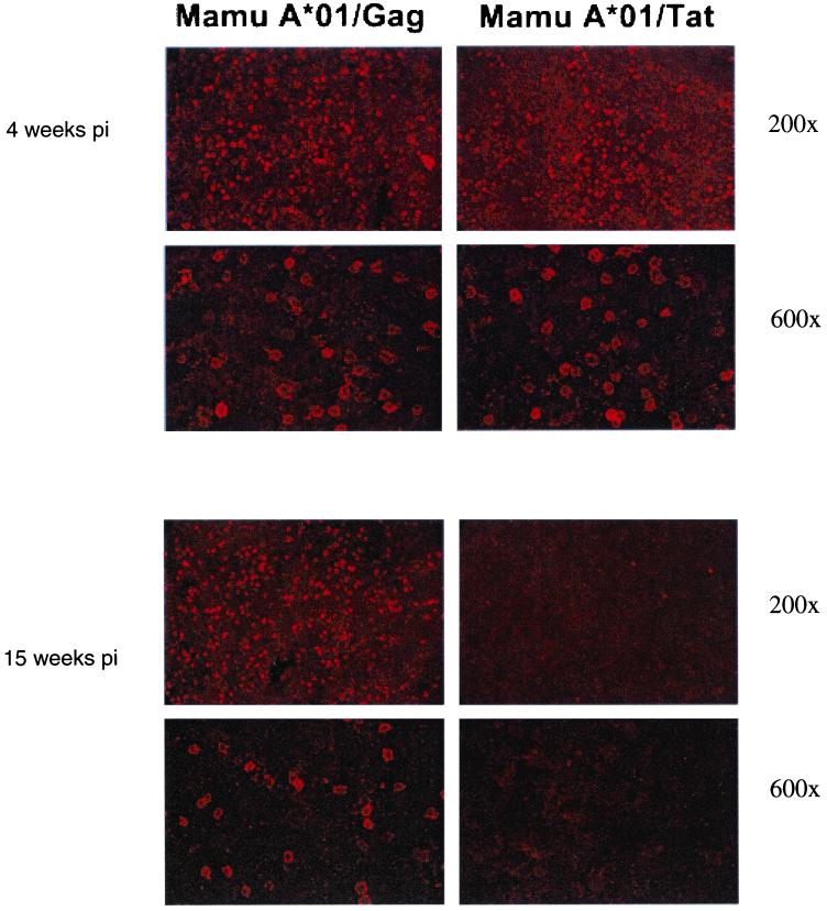
Cytotoxic T-lymphocyte (CTL) responses are thought to control human immunodeficiency virus replication during the acute phase of infection. Understanding the CD8(+) T-cell immune responses early after infection may, therefore, be important to vaccine design. Analyzing these responses in humans is difficult since few patients are diagnosed during early infection. Additionally, patients are infected by a variety of viral subtypes, making it hard to design reagents to measure their acute-phase immune responses. Given the complexities in evaluating acute-phase CD8(+) responses in humans, we analyzed these important immune responses in rhesus macaques expressing a common rhesus macaque major histocompatibility complex class I molecule (Mamu-A*01) for which we had developed a variety of immunological assays. We infected eight Mamu-A*01-positive macaques and five Mamu-A*01-negative macaques with the molecularly cloned virus SIV(mac)239 and determined all of the simian immunodeficiency virus-specific CD8(+) T-cell responses against overlapping peptides spanning the entire virus. We also monitored the evolution of particular CD8(+) T-cell responses by tetramer staining of peripheral lymphocytes as well as lymph node cells in situ. In this first analysis of the entire CD8(+) immune response to autologous virus we show that between 2 and 12 responses are detected during the acute phase in each animal. CTL against the early proteins (Tat, Rev, and Nef) and against regulatory proteins Vif and Vpr dominated the acute phase. Interestingly, CD8(+) responses against Mamu-A*01-restricted epitopes Tat(28-35)SL8 and Gag(181-189)CM9 were immunodominant in the acute phase. After the acute phase, however, this pattern of reactivity changed, and the Mamu-A*01-restricted response against the Gag(181-189)CM9 epitope became dominant. In most of the Mamu-A*01-positive macaques tested, CTL responses against epitopes bound by Mamu-A*01 dominated the CD8(+) cellular immune response.


Figures

FIG. 1.
FIG. 1.
Pools of peptides spanning all the proteins from SIVmac239. Our peptide set corresponds exactly to the sequence of the molecularly cloned SIVmac239 that we used for infection (except for one set of Pol 20-mer peptides, which corresponds to the sequence of SIVmac251 and is 99% identical to that of SIVmac239). Pools of peptides are used for intracellular cytokine staining for IFN-γ to determine the entire repertoire of CD8 antigen-specific responses.
FIG. 2.
FIG. 2.
Detection of IFN-γ production using the intracellular cytokine staining assay. PBMC from Mamu-A*01-positive SIV-infected animal 95096 were tested with Gag E and Tat A pools, the Gag 46 and Tat 12 15-mer peptides (pep), and the Gag181-189CM9 and Tat28-35SL8 peptides. Results depict the production of CD69 and IFN-γ in response to each pool of peptides and peptides for CD8+ lymphocytes. PMA, phorbol myristate acetate; Iono, ionomycin.
FIG. 3.
FIG. 3.
Acute-phase CD8+ T-cell responses in Mamu-A*01-positive macaques. Intracellular cytokine staining was performed on lymphocytes using pools spanning all the SIV proteins during the acute phase of SIVmac239 infection (3 to 4 weeks postinfection) in eight Mamu-A*01-positive animals. Bars, percentages of CD8+ IFN-γ+ responses for every SIV protein in each animal; last bar on the right, average percent contribution of each protein to the overall CD8+ response. Responses to the Gag and Tat proteins predominate in the acute phase of infection in most Mamu-A*01-positive animals. Number above each bar, number of responses detected in the animal.
FIG. 4.
FIG. 4.
Contribution of CD8+ responses directed against pools containing Mamu-A*01-bound peptides to acute-phase responses. Intracellular cytokine staining was performed on lymphocytes using pools spanning all the SIV proteins and Mamu-A*01 epitopes during the acute phase of SIVmac239 infection (3 to 4 weeks postinfection) in eight Mamu-A*01-positive animals. Bars, percentages of CD8+ IFN-γ+ responses for each SIV protein (except the Tat A and the Gag E pools) and the pools containing the Mamu-A*01-restricted epitopes, the Gag181-189CM9 and Tat28-35SL8 epitopes, in each animal. Responses to the pools containing the Mamu-A*01 epitopes predominate in the acute phase of infection in most Mamu-A*01-positive animals.
FIG. 5.
FIG. 5.
Tat28-35SL8 responses predominate in Mamu-A*01-positive rhesus macaques in the acute phase of SIV infection as visualized by tetramer staining of fresh PBMC. Ten Mamu-A*01-positive rhesus macaques were infected with cloned SIVmac239. Fresh PBMC were stained for CD3 and CD8 and the specific tetramer at room temperature. CD8+ responses to the two predominant Mamu-A*01-restricted epitopes, Gag181-189CM9 and Tat28-35SL8, were monitored using tetramers specific for each epitope. In the first few weeks postinfection the Tat28-35SL8-specific response predominates.
FIG. 6.
FIG. 6.
Gag- and Tat-specific T cells in lymph nodes from acute and chronic SIV infection by in situ tetramer staining. Images show lymph node sections from acutely and chronically SIV-infected macaques stained with Mamu-A*01-Gag181-189CM9- or Mamu-A*01-Tat28-35SL8-specific tetramers. Note the loss of detection of Tat-specific T cells in the sections from a chronically infected animal. Sections were counterstained with CD8+ antibodies (not shown). Each ×200 image is a projection of nine confocal Z-scans collected at 2-μm intervals. Each of the ×600 images was made by projecting 14 confocal Z-scans collected at 1-μm intervals.
FIG. 7.
FIG. 7.
Acute-phase CD8+ T-cell responses in Mamu-A*01-negative macaques. Intracellular cytokine staining was performed on lymphocytes using pools spanning all the SIV proteins during the acute phase of SIVmac239 infection (3 weeks postinfection) in five Mamu-A*01-negative animals. Bars, percentages of CD8+ IFN-γ+ responses for every SIV protein in each animal; last bar on the right, average percent contribution of each protein to the overall CD8+ response; number above each bar, number of responses detected in the animal.
FIG. 8.
FIG. 8.
Relative contribution of each protein to CD8 responses in the acute phase in all animals tested using ICS. (A) Total IFN-γ response to each protein per 100 amino acids. The sum of the IFN-γ responses to each protein was calculated and divided by the amino acid length of each protein. This number was then multiplied by 100 to determine the contribution per 100 amino acids. Additionally, we calculated the relative contribution of each protein without the pools containing the Gag181-189CM9 and Tat28-35SL8 responses. (B) Relative number of responses to each protein detected per 100 amino acids. The sum of the number of responses to each protein was calculated and divided by the amino acid length of each protein. This number was then multiplied by 100 to determine the contribution per 100 amino acids. Additionally, we calculated the relative contribution of each protein without the pools containing the Gag181-189CM9 and Tat28-35SL8 responses. Regulatory and accessory proteins Tat, Rev, Nef, and Vif were best recognized. On the other hand, structural proteins Env, Gag, and Pol were poorly recognized.

References

    1. Addo, M. M., M. Altfeld, E. S. Rosenberg, R. L. Eldridge, M. N. Philips, K. Habeeb, A. Khatri, C. Brander, G. K. Robbins, G. P. Mazzara, P. J. Goulder, and B. D. Walker. 2001. The HIV-1 regulatory proteins Tat and Rev are frequently targeted by cytotoxic T lymphocytes derived from HIV-1-infected individuals Proc. Natl. Acad. Sci. USA 98:1781–1786. - PMC - PubMed
    1. Allen, T. M., D. H. O’Connor, P. Jing, J. L. Dzuris, B. R. Mothe, E. Dunphy, M. E. Liebl, T. U. Vogel, C. Emerson, N. Wilson, K. J. Kunstman, X. Wang, A. L. Hughes, R. C. Desrosiers, J. D. Altman, S. M. Wolinsky, A. Sette, and D. I. Watkins. 2000. Tat-specific CTL select for SIV escape variants during resolution of primary viremia. Nature 407:386–390. - PubMed
    1. Altfeld, M. A., A. Trocha, R. L. Eldridge, E. S. Rosenberg, M. N. Phillips, M. M. Addo, R. P. Sekaly, S. A. Kalams, S. A. Burchett, K. McIntosh, B. D. Walker, and P. J. Goulder. 2000. Identification of dominant optimal HLA-B60- and HLA-B61-restricted cytotoxic T-lymphocyte (CTL) epitopes: rapid characterization of CTL responses by enzyme-linked immunospot assay. J. Virol. 74:8541–8549. - PMC - PubMed
    1. Amara, R. R., F. Villinger, J. D. Altman, S. L. Lydy, S. P. O’Neil, S. J. Staprans, D. C. Montefiori, Y. Xu, J. G. Herndon, L. S. Wyatt, M. A. Candido, N. L. Kozyr, P. L. Earl, J. M. Smith, H.-L. Ma, B. D. Grimm, M. L. Hulsey, H. M. McClure, J. M. McNicholl, B. Moss, and H. L. Robinson. 2001. Control of a mucosal challenge and prevention of AIDS by a multiprotein DNA/MVA vaccine. Science 292:69–74. - PubMed
    1. Barouch, D. H., S. Santra, J. E. Schmitz, M. J. Kuroda, T. M. Fu, W. Wagner, M. Bilska, A. Craiu, X. X. Zheng, G. R. Krivulka, K. Beaudry, M. A. Lifton, C. E. Nickerson, W. L. Trigona, K. Punt, D. C. Freed, L. Guan, S. Dubey, D. Casimiro, A. Simon, M. E. Davies, M. Chastain, T. B. Strom, R. S. Gelman, D. C. Montefiori, and M. G. Lewis. 2000. Control of viremia and prevention of clinical AIDS in rhesus monkeys by cytokine-augmented DNA vaccination. Science 290:486–492. - PubMed

Publication types

MeSH terms