. 2002 Jan;76(2):895-904.
doi: 10.1128/jvi.76.2.895-904.2002.

Genetically engineered vesicular stomatitis virus in gene therapy: application for treatment of malignant disease

Affiliations

Genetically engineered vesicular stomatitis virus in gene therapy: application for treatment of malignant disease

Marilyn Fernandez et al. J Virol. 2002 Jan.

Abstract

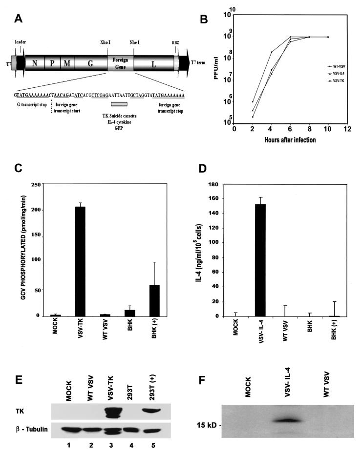
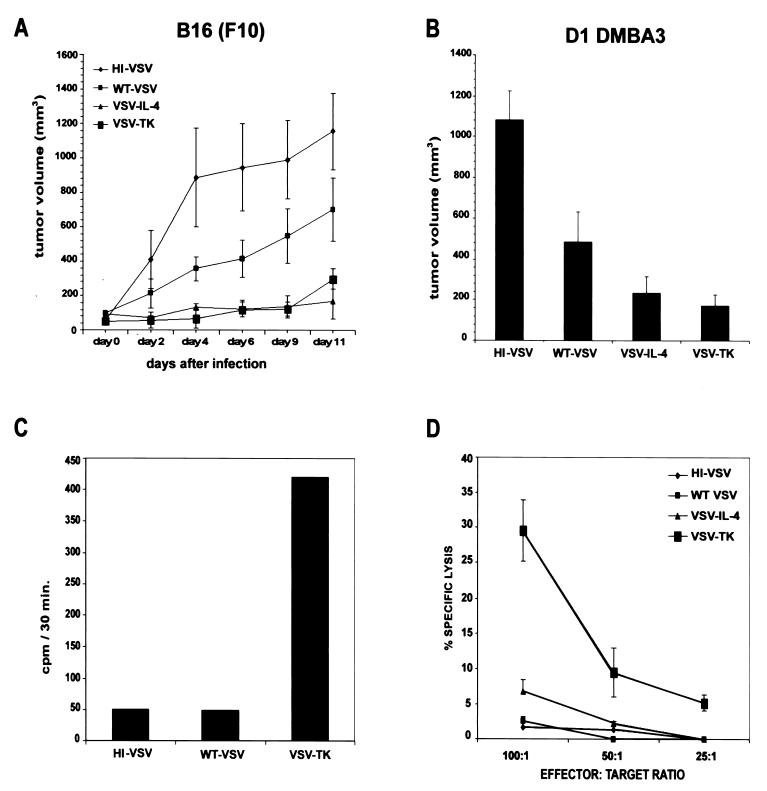
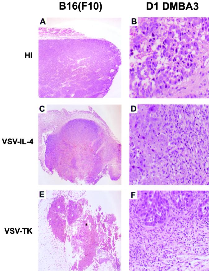
We report here the generation of recombinant vesicular stomatitis virus (VSV) able to produce the suicide gene product thymidine kinase (TK) or cytokine interleukin 4 (IL-4). In vitro cells infected with the engineered viruses expressed remarkably high levels of biologically active TK or IL-4 and showed no defects in replication compared to the wild-type virus. Recombinant viruses retained their ability to induce potent apoptosis in a variety of cancer cells, while normal cells were evidently more resistant to infection and were completely protected by interferon. Significantly, following direct intratumoral inoculation, VSV expressing either TK or IL-4 exhibited considerably more oncolytic activity against syngeneic breast or melanoma tumors in murine models than did the wild-type virus or control recombinant viruses expressing green fluorescent protein (GFP). Complete regression of a number of tumors was achieved, and increased granulocyte-infiltrating activity with concomitant, antitumor cytotoxic T-cell responses was observed. Aside from discovering greater oncolytic activity following direct intratumoral inoculation, however, we also established that VSV expressing IL-4 or TK, but not GFP, was able to exert enhanced antitumor activity against metastatic disease. Following intravenous administration of the recombinant viruses, immunocompetent BALB/c mice inoculated with mammary adenocarcinoma exhibited prolonged survival against lethal lung metastasis. Our data demonstrate the validity of developing novel types of engineered VSV for recombinant protein production and as a gene therapy vector for the treatment of malignant and other disease.


Figures

FIG. 1.
FIG. 1.
Generation of rVSV expressing TK, IL-4, or GFP. (A) cDNA representing the VSV genome (pVSV-XN2), flanked by the T7 RNA polymerase leader and T7 terminator as well as hepatitis virus delta ribozyme (RBZ), was used to create recombinant viruses. IL-4, TK, or GFP was inserted between the glycoprotein and polymerase protein genes of VSV. (B) Growth curves of recombinant viruses. BHK cells were infected with wild-type VSV, VSV-IL-4, and VSV-TK at an MOI of 10. Supernatants from infected cells were harvested at the indicated times postinfection, and viral titers were determined by plaque assay. (C) GCV is phosphorylated in cells infected with VSV-TK. BHK cells were mock infected or infected with VSV-TK or wild-type (WT) VSV (MOI = 1) for 8 h, and cell lysates were assayed for GCV phosphorylation in vitro. BHK cells transiently transfected with CMV promoter-driven HSV-TK [BHK(+)] or empty vector (BHK) were used as controls. (D) High-level expression of IL-4 in cells infected with VSV-IL-4. Culture medium from BHK cells infected with wild-type VSV or VSV-IL-4 was measured for functional IL-4 using capture ELISA. As a further control, IL-4 was measured in culture medium from BHK cells transiently transfected with an empty vector or with CMV promoter-driven IL-4 cDNA. (E) Expression of HSV-TK in VSV-TK-infected cells. BHK cells were mock infected (lane 1) or infected with wild-type VSV (lane 2) or VSV-TK (lane 3) at an MOI of 1 for 24 h, and cell extracts were analyzed for TK expression using an anti-TK monoclonal antibody. 293T cells transiently transfected with an empty vector (lane 4) or with CMV promoter-driven HSV-TK (lane 5) were used as a positive control. (F) Immunoprecipitation of IL-4 from supernatants of VSV-IL-4-infected cells. Extracts from [35S]methionine-labeled cells mock infected (lane 1) or infected with VSV-IL-4 (lane 2) or wild-type VSV (lane 3) were immunoprecipitated with an IL-4 antibody.
FIG. 2.
FIG. 2.
In vitro effects of wild-type (WT) VSV and rVSV on primary or transformed cells. (A to C) rVSVs efficiently kill transformed cells. HMVECs or B16(F10) or DA-3 cells were infected with wild-type VSV, VSV-TK, or VSV-IL-4 with (solid columns) or without (empty columns) prior treatment with IFN-α. Cell viability was assayed by trypan blue exclusion 18 h after infection. (D to F) Efficient replication of VSV-GFP in transformed cells. HMVECs or B16(F10) or DA-3 cells were infected with VSV-GFP with or without prior treatment of IFN-α (1,000 U/ml). Top panels show cells under bright field microscopy (magnification, ×20), and lower panels show the same field by immunofluorescence.
FIG. 3.
FIG. 3.
rVSV expressing TK and IL-4 inhibits the growth of syngeneic breast and melanoma tumors in immunocompetent mice. (A) C57BL/6 mice were implanted subcutaneously with 5 × 10⁵ B16(F10) melanoma cells. After palpable tumors had formed, animals were treated intratumorally with 2 × 10⁷ PFU of wild-type VSV, VSV-IL-4, or VSV-TK. Injections of virus were repeated after 3 days. Tumor volumes were calculated and are shown as a mean ± standard error of the mean (n = 5). Two mice that received heat-inactivated virus were sacrificed at day 4 due to the large size of tumors. (B) BALB/c mice were implanted subcutaneously with 1.5 × 10⁶ D1-DMBA3 tumor cells. After palpable tumors had formed, animals were intratumorally injected with 2 × 10⁷ PFU of heat-inactivated (HI) virus, wild-type VSV, VSV-IL-4, or VSV-TK. Virus treatment was repeated after 3 days. Tumor volumes at day 21 postimplantation (7 days after the last virus treatment) are shown. Results are presented as a mean ± standard error of the mean (n = 5). Comparable results were obtained in three independent sets of experiments. (C) GCV phosphorylation in vivo in B16(F10) tumor cells following VSV-TK administration. B16(F10) tumors were harvested from C57BL/6 mice 1 day following intratumoral injection with 2 × 10⁷ PFU of heat-inactivated VSV, wild-type VSV, or VSV-TK. The amount of GCV phosphorylated was measured in extracts (60 μg of total protein) from tumor cells 30 min following incubation in a reaction mixture containing [3H]GCV (45 μM). (D) Induction of CTL response against B16(F10) tumors in animals receiving VSV-TK-GCV treatment. C57BL/6 tumor-bearing mice were injected intratumorally with wild-type VSV or rVSV. A second injection was administered 3 days later. Ten days after the first virus injection, spleen cells were isolated and cocultured with B16(F10) cells. Spleen cells were incubated at the indicated effector-to-target ratios with 51Cr-labeled B16(F10) target cells. CTL activity was determined by 51Cr release.
FIG. 4.
FIG. 4.
Histopathological analysis of tumors. Tumors from C57BL/6 and BALB/c mice were removed 7 days after receiving intratumoral injections of heat-inactivated VSV (A), VSV-IL-4 (B), or VSV-TK (C). The left panels indicate large areas of cell death in B16(F10) tumors treated with VSV-TK and VSV-IL-4. The right panels emphasize increased infiltration of eosinophils in D1-DMBA3 tumors injected with VSV-IL-4.
FIG. 5.
FIG. 5.
Intravenous treatment with VSV-TK and VSV-IL-4 increases the survival of mice in a metastatic tumor model. (A) Effects of rVSV on TS/A cells in vitro. TS/A cells were treated with or without murine IFN (1,000 U/ml) for 24 h and were infected with rVSV at an MOI of 0.1 for 18 h. Cell viability in response to virus infection was measured using trypan blue. (B) Effects of rVSVs against metastatic disease. BALB/c mice (n = 10 per group) were injected via the tail vein with 5 × 10⁴ TS/A cells, followed 4 days later by intravenous administration of 5 × 10⁶ PFU of either heat-inactivated (HI) VSV, VSV-GFP, VSV-TK, or VSV-IL-4. The survival rate of mice following virus injection is shown. (C) Morphological examination of TS/A cells following rVSV infection in vitro.

Similar articles

Cited by

References

    1. Asnagli, H., and K. M. Murphy. 2001. Stability and commitment in T helper cell development. Curr. Opin. Immunol. 13:242–247. - PubMed
    1. Balachandran, S., and G. N. Barber. 2000. Vesicular stomatitis virus (VSV) therapy of tumors. IUBMB Life 50:135–138. - PubMed
    1. Balachandran, S., M. Porosnicu, and G. N. Barber. 2001. Oncolytic activity of vesicular stomatitis virus is effective against tumors exhibiting aberrant p53, Ras, or Myc function and involves the induction of apoptosis. J. Virol. 75:3474–3479. - PMC - PubMed
    1. Balachandran, S., P. C. Roberts, L. E. Brown, H. Truong, A. K. Pattnaik, D. R. Archer, and G. N. Barber. 2000. Essential role for the dsRNA-dependent protein kinase PKR in innate immunity to viral infection. Immunity 13:129–141. - PubMed
    1. Ball, L. A., C. R. Pringle, B. Flanagan, V. P. Perepelitsa, and G. W. Wertz. 1999. Phenotypic consequences of rearranging the P, M, and G genes of vesicular stomatitis virus. J. Virol. 73:4705–4712. - PMC - PubMed

MeSH terms