

CDD: a database of conserved domain alignments with links to domain three-dimensional structure
- PMID: 11752315
- PMCID: PMC99109
- DOI: 10.1093/nar/30.1.281
CDD: a database of conserved domain alignments with links to domain three-dimensional structure
Abstract
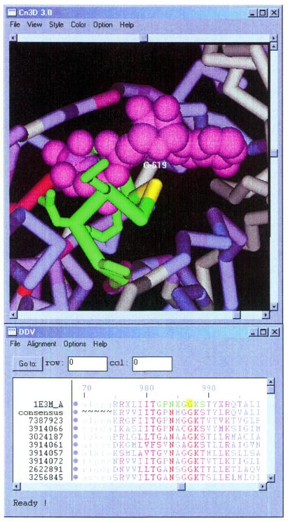
The Conserved Domain Database (CDD) is a compilation of multiple sequence alignments representing protein domains conserved in molecular evolution. It has been populated with alignment data from the public collections Pfam and SMART, as well as with contributions from colleagues at NCBI. The current version of CDD (v.1.54) contains 3693 such models. CDD alignments are linked to protein sequence and structure data in Entrez. The molecular structure viewer Cn3D serves as a tool to interactively visualize alignments and three-dimensional structure, and to link three-dimensional residue coordinates to descriptions of evolutionary conservation. CDD can be accessed on the World Wide Web at http://www.ncbi.nlm.nih.gov/Structure/cdd/cdd.shtml. Protein query sequences may be compared against databases of position-specific score matrices derived from alignments in CDD, using a service named CD-Search, which can be found at http://www.ncbi.nlm.nih.gov/Structure/cdd/wrpsb.cgi. CD-Search runs reverse-position-specific BLAST (RPS-BLAST), a variant of the widely used PSI-BLAST algorithm. CD-Search is run by default for protein-protein queries submitted to NCBI's BLAST service at http://www.ncbi.nlm.nih.gov/BLAST.
Figures
References
-
- Park J., Karplus,K., Barrett,C., Hughey,R., Haussler,D., Hubbard,T. and Chothia,C. (1998) Sequence comparisons using multiple sequences detect three times as many remote homologues as pairwise methods. J. Mol. Biol., 284, 1201–1210. - PubMed
-
- Eddy S.R. (1998) Profile hidden Markov models. Bioinformatics, 14, 755–763. - PubMed
-
- Karplus K., Barrett,C. and Hughey,R. (1998) Hidden Markov models for detecting remote protein homologies. Bioinformatics, 14, 846–856. - PubMed
MeSH terms
Substances
LinkOut - more resources
Full Text Sources
Other Literature Sources
Research Materials
