. 2001 Dec;13(12):2731-45.
doi: 10.1105/tpc.010253.

Functional studies of Ycf3: its role in assembly of photosystem I and interactions with some of its subunits

Affiliations

Functional studies of Ycf3: its role in assembly of photosystem I and interactions with some of its subunits

H Naver et al. Plant Cell. 2001 Dec.

Abstract

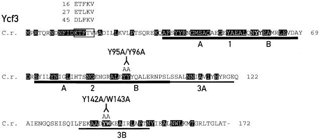
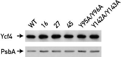
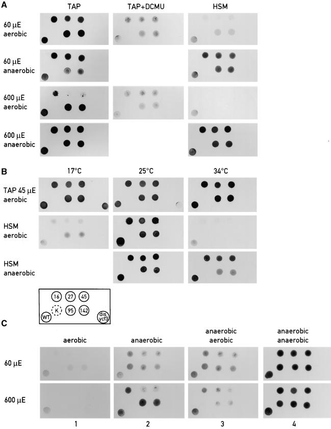
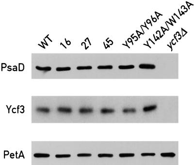
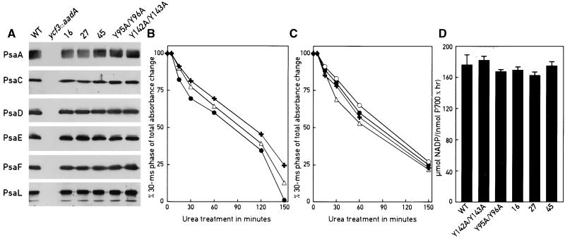
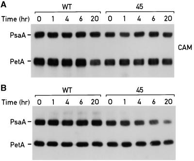
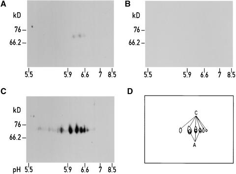
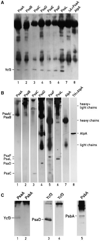
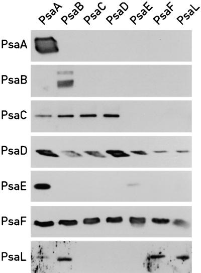
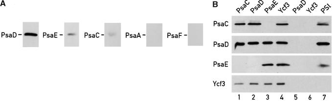
The Ycf3 protein is essential for the accumulation of the photosystem I (PSI) complex and acts at a post-translational level. The sequence of Ycf3 is conserved in cyanobacteria, algae, and plants and contains three tetratrico-peptide repeats (TPR). TPRs have been shown to function as sites for protein-protein interactions. The mutations Y95A/Y96A and Y142A/W143A in the second and third TPR repeats lead to a modest decrease of PSI, but they prevent photoautotrophic growth and cause enhanced light sensitivity even though the accumulated PSI complex is fully functional. This phenotype can be reversed under anaerobic conditions and appears to be the result of photooxidative damage. A temperature-sensitive ycf3 mutant, generated by random mutagenesis of a conserved region near the N-terminal end of Ycf3, was used in temperature-shift experiments to show that Ycf3 is required for PSI assembly but not for its stability. Immunoblot analysis of thylakoid membranes separated by two-dimensional gel electrophoresis and immunoprecipitations shows that Ycf3 interacts directly with the PSI subunits PsaA and PsaD, but not with subunits from other photosynthetic complexes. Thus, Ycf3 appears to act as a chaperone that interacts directly and specifically with at least two of the PSI subunits during assembly of the PSI complex.


Figures

Figure 1.
Figure 1.
Mutations within ycf3. The sequence of Ycf3 from C. reinhardtii (C.r.) is shown. Residues conserved in Ycf3 from liverwort, tobacco, black pine, Odontella sinensis, Cyanidium caldarium, Porphyra purpurea, Cyanophora paradoxa, and Synechocstis PCC 6803 are shaded. The region subjected to degenerate oligonucelotide mutagenesis is boxed, and the changes in the mutants 16, 27, and 45 are indicated. The C. reinhardtii TPR motifs are underlined, and the changes in the TPR domains 2 (Y95A/Y96A) and 3 (Y142A/W143A) are indicated. The regions corresponding to the TPR subdomains A and B are marked, and the borders between the subdomains are indicated with arrowheads.
Figure 2.
Figure 2.
Ycf4 Accumulates to Wild-Type Levels in Selected ycf3 Mutants. Thylakoid protein (10 μg) from the wild type (WT) and ycf3 mutants were separated on a 12% polyacrylamide gel and probed with antibodies against Ycf4 and PsbA. Equal loading of proteins was tested by probing the blot with PsbA antibody.
Figure 3.
Figure 3.
Growth Patterns of the Wild Type and ycf3 Mutant Strains. (A) Photosensitivity of ycf3 mutants. Cells were plated on TAP, TAP with 5 μM DCMU, or HSM plates and grown at light intensities of 60 or 600 μE·m−2·sec−1 under aerobic and anaerobic conditions. (B) Temperature sensitivity of ycf3 mutants. Cells were plated on either TAP or HSM plates and grown at 17, 25, or 34°C at a light intensity of 45 μE·m−2·sec−1. Mutants 16, 27, and 45 are described in Figure 1. The arrangement of the mutants and wild type are indicated in the block scheme. Mutant 95, Y95A/Y96A; mutant 142, Y142A/W143A; dis.ycf3, ycf3::aadA disruption mutant; WT, wild type. (C) Anaerobic–aerobic transfers. The same mutant cells were used except that the psaC mutant K35E was added (in [B], indicated by K surrounded by a broken circle). Cells were grown for 3 days at 60 or 600 μE·m−2·sec−1 under aerobic (lane 1) or anaerobic (lane 2) conditions. Cells growing anaerobically were then grown aerobically (lane 3) or maintained under anaerobic conditions (lane 4) for 4 additional days.
Figure 4.
Figure 4.
PSI Accumulation in the ycf3 Mutants. Total cell proteins (40 μg) from the wild type (WT) and the ycf3 mutants were separated on a 12% polyacrylamide gel, immunoblotted, and probed with antibodies against PsaD, Ycf3, and PetA (which was used as a loading control).
Figure 5.
Figure 5.
Composition and Functional Stability of the PSI Complex from the ycf3 Mutants and the Wild Type Are Undistinguishable. (A) PSI was purified from each of the mutant strains 16, 27, 45 (see Figure 1 for description), 95 (Y95A/Y96A), 142 (Y142A/W143A), and the wild type (WT). In the case of ycf3::aadA, the thylakoid membrane fraction corresponding to PSI in the wild type was used. Aliquots (5 μg of protein) of each preparation were loaded onto SDS–polyacrylamide gels and analyzed by immunoblotting with the antibodies indicated. (B) and (C) Stability of PSI in the presence of 6.8 M urea. PSI preparations from the wild type, 16, 27, 45, Y95A/Y96A, and Y142A/W143A were analyzed by flash-induced absorption transients at 817 nm after incubation with urea. Charge recombination between electron acceptors in the PSI complex [FA/FB] , FX, A1, and P700+ was recorded. Before urea treatment, charge recombination is monophasic, consisting exclusively of the [FA/FB] to P700+ charge recombination of >30 msec. After addition of urea, charge recombination becomes biphasic, consisting of recombinations between [FA/FB] and P700+ and between FX and P700+. The proportion of the absorbance change corresponding to the 30-msec phase ([FA/FB] and P700+) relative to the total absorbance change is shown. In (B), black crosses, Y95A/Y96A; open triangles, wild type; solid circles, Y142A/W143A. In (C), open triangles, 27; crosses, 16; closed circles, wild type; open circles, 45. (D) In vitro PSI activity of the wild type (WT) and the mutant strains. NADP+ photo reduction of PSI from the indicated strains was measured on isolated PSI complexes as described in Methods. Error bars indicte ±sd.
Figure 6.
Figure 6.
Ycf3 Is Required for PSI Assembly but Not for Its Stability. Cultures from the wild type (WT) and the ycf3 mutant 45 were grown in 50 mL of TAP at 18°C at a light intensity of 45 μE·m−2·sec−1 until the cell density reached 2 × 10⁶ cells mL−1. Cells were pelleted by centrifugation, washed in HSM, and resuspended to the same concentration in HSM. Each culture was split into two parts. Both cultures were incubated for 2 hr at 18°C at a light intensity of 45 μE·m−2·sec−1 and shifted to 34°C at the same light intensity. (A) Chloramphenicol (CAM; 200 μg·mL−1) was added to one culture at the same time as the temperature shift to inhibit chloroplast protein synthesis. Samples of 150 μL were transferred to 1.5 mL of cold acetone (−20°C) at 0, 1, 4, 6, and 20 hr after the shift to 34°C. The acetone-precipitated cells were boiled in sample buffer, loaded on a 12% SDS–polyacrylamide gel, and immunoblotted. (B) Samples (1 mL) were collected from the other culture at 0, 1, 4, 6 and 20 hr after the shift to 34°C. Samples were solubilized, the protein concentrations were measured, and sample volumes corresponding to 40 μg of protein were boiled in sample buffer, loaded onto 12% SDS–polyacryamide gels, immunoblotted, and probed with antibodies against PetA and PsaA.
Figure 7.
Figure 7.
Ycf3 Interacts with PsaA. Wild-type thylakoid membrane proteins (200 μg per gel) were separated by two-dimensional gel electrophoresis on four gels, which were each transferred to nitrocellulose membranes. (A) The nitrocellulose membrane was incubated with recombinant His-tagged Ycf3 protein and subsequently reacted with His antibodies. (B) After blotting, the membrane was reacted with His antibody. (C) After blotting, the membrane was reacted with PsaA antibody. (D) Superposition of (A) and (C).
Figure 8.
Figure 8.
Ycf3 Co-Immunoprecipitates with PsaA, PsaB, PsaC, PsaD, PsaE, PsaF, and PsaL. Solubilized thylakoid membranes from the wild-type strain were immunoprecipitated with antibodies cross-linked to cyanogen bromide–activated Sepharose 4B. The Sepharose beads were washed, and the immunoprecipitates eluted. The eluates were fractionated by 10 to 17% polyacrylamide gels and immunoblotted. (A) Immunoprecipitation with PsaA, PsaB, PsaC, PsaD, PsaE, PsaF, PsaL, and AtpA antibodies. Lane 7 marked ΔA+PsaA contains solubilized thylakoids, from the psaAΔ strain that accumulates wild-type levels of Ycf3 incubated with PsaA antibody. The immunoprecipitates were eluted with Laemmli SDS sample buffer, immunoblotted, and probed with Ycf3 antibody. The antibodies used for the immunoprecipitation are indicated at the top of each lane. (B) Immunoprecipitation with Ycf3 antibody. The immunoprecipitates were eluted with 0.1 M glycine, pH 2.8 (lanes 1, to 3), with Laemmli SDS sample buffer (lanes 4, 5, and 7), or with Laemmli SDS sample buffer without DTT (lane 6; under these conditions the IgG heavy and light chains do not dissociate). Lane 8 shows an immunoblot of thylakoid membrane proteins reacted with AtpA antibodies. Lanes 1 to 4, 5 to 7, and 8 are from separate gels. The antibodies used for the immunoblots are indicated at the top of each lane. Heavy and light chains refer to the IgG chains. (C) Immunoprecipitation with PsaA, PsbA (D1), and Ycf3 antibodies. The antibodies used for the immunoprecipitation are indicated at the top of each lane. The immunoblots were probed with Ycf3 (lanes 1 and 2), PsaD (lane 3), and PsbA (lanes 4 and 5) antibodies as indicated.
Figure 9.
Figure 9.
PSI Subunit Content in Immunoprecipitations with PSI Antibodies. Thylakoid membranes from the wild-type strain were solubilized with 0.9% dodecylmaltoside at a chlorophyll concentration of 0.8 mg·mL−1 and immunoprecipitated with antibodies cross-linked to cyanogen bromide–activated Sepharose 4B. The Sepharose beads were washed, and the immunoprecipitates were eluted with 100 mM glycine, pH 2.8. The eluates were fractionated by 10 to 17% polyacrylamide gels and immunoblotted. The antibodies used for the immunoprecipitations are indicated at the top of each lane. The antibodies used to probe the immunoblots are indicated at left.
Figure 10.
Figure 10.
Ycf3 Interacts with PsaD. PSI preparations were extracted with butanol. The butanol phase containing the membrane proteins of PSI was discarded, and the water phase containing the soluble stromal subunits PsaC, PsaD, and PsaE was saved. (A) The PsaC, PsaD, and PsaE-enriched extract was fractionated on SDS–polyacrylamide gels, immunoblotted, and probed with PsaA, PsaC, PsaD, PsaE, and PsaF antibodies. (B) Immunoprecipitation of the PsaC, PsaD, and PsaE-enriched extract. The antibodies used for immunoprecipitation are indicated at the top of the lanes. The antibodies used to probe the immunoblots are indicated at left. Samples were immunoprecipitated with antibodies cross-linked to cyanogen bromide–activated Sepharose 4B. The Sepharose beads were washed, and the immunoprecipitates were eluted with 100 mM glycine, pH 2.8. The eluates were fractionated by 10 to 17% polyacrylamide gels and immunoblotted. Lanes 1 to 4, the PsaC, PsaD, and PsaE-enriched extract was mixed with thylakoid membranes solubilized with 0.9% dodecylmaltoside at a chlorophyll concentration of 0.8 mg·mL−1 from the psaA-deficient mutant ΔA containing Ycf3 in wild-type amounts and immunoprecipitated as indicated; lane 5, solubilized thylakoid membranes from the ΔA mutant were immunoprecipitated with PsaD antibody in the absence of the psaC, PsaD, and PsaE-enriched extract; lane 6, solubilized thylakoid membranes from the ycf3::aadA strain were mixed with the the PsaC, PsaD, and PsaE-enriched extract and immunoprecipitated with Ycf3 antibody; lane 7, purified PSI complex was used for immunoprecipitation.

Similar articles

Cited by

References

    1. Asada, K. (1994). Mechanisms for scavenging reactive molecules generated in chloroplasts under light stress. In Photoinhibition of Photosynthesis: From Molecular Mechanisms to the Field, N.R. Baker and J.R. Bowyer, eds (Oxford, UK: BIOS Scientific Publishers), pp. 129–142.
    1. Asada, K. (1996). Radical production and scavenging in the chloroplast. In Advances in Photosynthesis, N.R. Baker, ed (Dordrecht, The Netherlands: Kluwer Academic Publishers), pp. 123–150.
    1. Barber, J., and Andersson, B. (1992). Too much of a good thing: Light can be bad for photosynthesis. Trends Biochem. Sci. 17, 61–66. - PubMed
    1. Bartsevich, V.V., and Pakrasi, H.B. (1997). Molecular identification of a novel protein that regulates biogenesis of photosystem I, a membrane protein complex. J. Biol. Chem. 272, 6382–6387. - PubMed
    1. Bassi, R., Soen, S.Y., Frank, G., Zuber, H., and Rochaix, J.-D. (1992). Characterization of chlorophyll a/b proteins of photosystem I from Chlamydomonas reinhardtii. J. Biol. Chem. 267, 25714–25721. - PubMed

Publication types

Substances

LinkOut - more resources