. 2002 Jan 8;99(1):19-24.
doi: 10.1073/pnas.012583299. Epub 2001 Dec 18.

Incorporation of azides into recombinant proteins for chemoselective modification by the Staudinger ligation

Affiliations

Incorporation of azides into recombinant proteins for chemoselective modification by the Staudinger ligation

Kristi L Kiick et al. Proc Natl Acad Sci U S A. .

Abstract

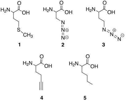
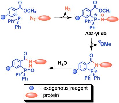
The introduction of chemically unique groups into proteins by means of non-natural amino acids has numerous applications in protein engineering and functional studies. One method to achieve this involves the utilization of a non-natural amino acid by the cell's native translational apparatus. Here we demonstrate that a methionine surrogate, azidohomoalanine, is activated by the methionyl-tRNA synthetase of Escherichia coli and replaces methionine in proteins expressed in methionine-depleted bacterial cultures. We further show that proteins containing azidohomoalanine can be selectively modified in the presence of other cellular proteins by means of Staudinger ligation with triarylphosphine reagents. Incorporation of azide-functionalized amino acids into proteins in vivo provides opportunities for protein modification under native conditions and selective labeling of proteins in the intracellular environment.


Figures

Figure 1
Figure 1
Structures of methionine and the analogs tested for utility as methionine surrogates. (1) Methionine, (2) azidoalanine, (3) azidohomoalanine, (4) 2-amino-5-hexynoic acid, and (5) norleucine.
Scheme 1
Scheme 1
The Staudinger ligation between a protein containing azide functionalized amino acid side chains and a phosphine reagent.
Figure 2
Figure 2
Electron density maps (colored surfaces) and negative isopotential surfaces (meshes) for (a) methionine, (b) 2, (c) 3, and (d) 4. The electron density maps indicate electron-rich (red) and electron-poor (blue) regions of each molecule. For simplicity, the amino acid form is shown; this avoids representation of the highly extended isopotential surface of the carboxylate anion of the zwitterion and facilitates comparison of side-chain electronic structures.
Figure 3
Figure 3
SDS/PAGE analysis of the translational activity of azide-functionalized amino acids. The SDS/PAGE analysis was conducted on whole-cell lysates of CAG18491/pQE15/pREP4 cultures, supplemented with nothing (−Met), methionine (+Met), 2, or 3.
Figure 4
Figure 4
N-terminal sequencing results indicating occupancy of the initiator site in mDHFR produced in bacterial cultures supplemented with 3. N-terminal residues are shown for (a) DHFR-Met, (b) the free amino acid 3, and (c) DHFR-3, as determined by Edman degradation. pptu, piperidylphenylthiourea; diet, diethylphthalate.
Figure 5
Figure 5
Mass spectrometric analysis of a tryptic fragment of mDHFR-3. The tryptic digest of mDHFR-3 was analyzed by MALDI-MS; results are shown for a fragment with an expected mass of 2,146.4 Da. The observed peak at a mass of 2,141.1 Da is consistent with that expected for the fragment in which 3 replaces methionine. The peak at 2,115.1 Da may correspond to a fragment in which 3 has been reduced to 2,4-diaminobutyric acid.
Scheme 2
Scheme 2
Synthesis of triarylphosphine-FLAG conjugate 9.
Figure 6
Figure 6
Western blot analysis of the products of Staudinger ligation. (a) Purified protein (mDHFR-3 or mDHFR-Met) was used in the ligation, and the blot was labeled with anti-FLAG M2 mAb followed by HRP-rat anti-mouse IgG1. (b) Similar to a but labeled with India HisProbe-HRP. (c) Crude cell lysate (containing either mDHFR-3 or mDHFR-Met) was used in the ligation, and the blot was labeled with anti-FLAG M2 mAb followed by HRP-rat anti-mouse IgG1. (d) Similar to c but labeled with India HisProbe-HRP.

Similar articles

Cited by

References

    1. Dougherty D A. Curr Opin Chem Biol. 2000;4:645–652. - PubMed
    1. Marcaurelle L A, Bertozzi C R. Chem Eur J. 1999;5:1384–1390.
    1. Cotton G J, Muir T W. Chem Biol. 1999;6:R247–R256. - PubMed
    1. Cornish V W, Mendel D, Schultz P G. Angew Chem Int Ed Engl. 1995;34:621–633.
    1. Mendel D, Ellman J A, Chang Z, Veenstra D L, Kollman P A, Schultz P G. Science. 1992;256:1798–1802. - PubMed

Publication types