

Regulatory DNA required for vnd/NK-2 homeobox gene expression pattern in neuroblasts
- PMID: 11752402
- PMCID: PMC117523
- DOI: 10.1073/pnas.012584599
Regulatory DNA required for vnd/NK-2 homeobox gene expression pattern in neuroblasts
Erratum in
- Proc Natl Acad Sci U S A 2002 Jun 11;99(12):8458-9
Abstract
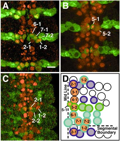
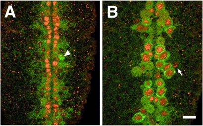
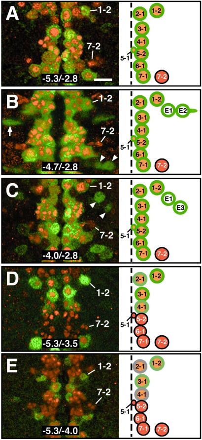
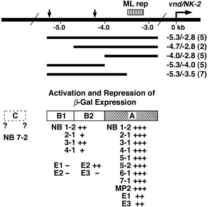
Vnd/NK-2 protein was detected in 11 neuroblasts per hemisegment in Drosophila embryos, 9 medial and 2 intermediate neuroblasts. Fragments of DNA from the 5'-flanking region of the vnd/NK-2 gene were inserted upstream of an enhancerless betagalactosidase gene in a P-element and used to generate transgenic fly lines. Antibodies directed against Vnd/NK-2 and beta-galactosidase proteins then were used in double-label experiments to correlate the expression of beta-galactosidase and Vnd/NK-2 proteins in identified neuroblasts. DNA region A, which corresponds to the -4.0 to -2.8-kb fragment of DNA from the 5'-flanking region of the vnd/NK-2 gene was shown to contain one or more strong enhancers required for expression of the vnd/NK-2 gene in ten neuroblasts. DNA region B (-5.3 to -4.0 kb) contains moderately strong enhancers for vnd/NK-2 gene expression in four neuroblasts. Hypothesized DNA region C, whose location was not identified, contains one or more enhancers that activate vnd/NK-2 gene expression only in one neuroblast. These results show that nucleotide sequences in at least three regions of DNA regulate the expression of the vnd/NK-2 gene, that the vnd/NK-2 gene can be activated in different ways in different neuroblasts, and that the pattern of vnd/NK-2 gene expression in neuroblasts of the ventral nerve cord is the sum of partial patterns.
Figures
Similar articles
-
Neuroblast pattern formation: regulatory DNA that confers the vnd/NK-2 homeobox gene pattern on a reporter gene in transgenic lines of Drosophila.Proc Natl Acad Sci U S A. 1998 Jul 7;95(14):8316-21. doi: 10.1073/pnas.95.14.8316. Proc Natl Acad Sci U S A. 1998. PMID: 9653184 Free PMC article.
-
Dorsal-ventral patterning genes restrict NK-2 homeobox gene expression to the ventral half of the central nervous system of Drosophila embryos.Dev Biol. 1995 Oct;171(2):306-16. doi: 10.1006/dbio.1995.1283. Dev Biol. 1995. PMID: 7556915
-
Formation and specification of ventral neuroblasts is controlled by vnd in Drosophila neurogenesis.Genes Dev. 1998 Nov 15;12(22):3613-24. doi: 10.1101/gad.12.22.3613. Genes Dev. 1998. PMID: 9832512 Free PMC article.
-
Unraveling cis-regulatory mechanisms at the abdominal-A and Abdominal-B genes in the Drosophila bithorax complex.Dev Biol. 2006 May 15;293(2):294-304. doi: 10.1016/j.ydbio.2006.02.015. Epub 2006 Mar 20. Dev Biol. 2006. PMID: 16545794 Review.
-
Molecular control of cell polarity and asymmetric cell division in Drosophila neuroblasts.Curr Opin Cell Biol. 2005 Oct;17(5):475-81. doi: 10.1016/j.ceb.2005.08.005. Curr Opin Cell Biol. 2005. PMID: 16099639 Review.
Cited by
-
Ind represses msh expression in the intermediate column of the Drosophila neuroectoderm, through direct interaction with upstream regulatory DNA.Dev Dyn. 2009 Nov;238(11):2735-44. doi: 10.1002/dvdy.22096. Dev Dyn. 2009. PMID: 19795518 Free PMC article.
-
cis-Decoder discovers constellations of conserved DNA sequences shared among tissue-specific enhancers.Genome Biol. 2007;8(5):R75. doi: 10.1186/gb-2007-8-5-r75. Genome Biol. 2007. PMID: 17490485 Free PMC article.
-
Genetic regulation and function of epidermal growth factor receptor signalling in patterning of the embryonic Drosophila brain.Open Biol. 2016 Dec;6(12):160202. doi: 10.1098/rsob.160202. Open Biol. 2016. PMID: 27974623 Free PMC article.
-
Evolution of insect dorsoventral patterning mechanisms.Cold Spring Harb Symp Quant Biol. 2009;74:275-9. doi: 10.1101/sqb.2009.74.021. Epub 2009 Oct 20. Cold Spring Harb Symp Quant Biol. 2009. PMID: 19843594 Free PMC article. Review.
-
Identification and analysis of vnd/NK-2 homeodomain binding sites in genomic DNA.Proc Natl Acad Sci U S A. 2005 May 17;102(20):7097-102. doi: 10.1073/pnas.0502261102. Epub 2005 May 3. Proc Natl Acad Sci U S A. 2005. PMID: 15870192 Free PMC article.
References
MeSH terms
Substances
LinkOut - more resources
Full Text Sources
Molecular Biology Databases
Research Materials
