. 2001 Dec 18;98(26):14790-5.
doi: 10.1073/pnas.221580098.

Structural basis for the activation of human procaspase-7

Affiliations

Structural basis for the activation of human procaspase-7

S J Riedl et al. Proc Natl Acad Sci U S A. .

Abstract

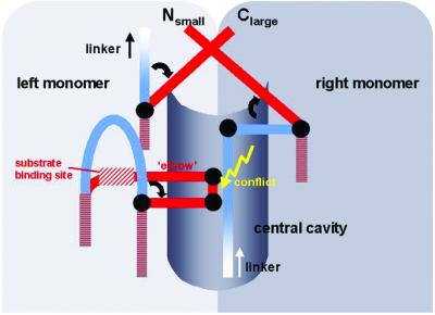
Caspases form a family of proteinases required for the initiation and execution phases of apoptosis. Distinct proapoptotic stimuli lead to activation of the initiator caspases-8 and -9, which in turn activate the common executioner caspases-3 and -7 by proteolytic cleavage. Whereas crystal structures of several active caspases have been reported, no three-dimensional structure of an uncleaved caspase zymogen is available so far. We have determined the 2.9-A crystal structure of recombinant human C285A procaspase-7 and have elucidated the activation mechanism of caspases. The overall fold of the homodimeric procaspase-7 resembles that of the active tetrameric caspase-7. Each monomer is organized in two structured subdomains connected by partially flexible linkers, which asymmetrically occupy and block the central cavity, a typical feature of active caspases. This blockage is incompatible with a functional substrate binding site/active site. After proteolytic cleavage within the flexible linkers, the newly formed chain termini leave the cavity and fold outward to form stable structures. These conformational changes are associated with the formation of an intact active-site cleft. Therefore, this mechanism represents a formerly unknown type of proteinase zymogen activation.


Figures

Figure 1
Figure 1
Integrity of crystallized human procaspase-7. Lane 1, molecular weight markers; lane 2, ≈8 μg of recombinant procaspase-7 used for crystallization trials; lane 3, recombinant procaspase-7 after treatment with granzyme B; lane 4, dissolved crystal of procaspase-7; lane 5, dissolved crystal of procaspase-7 treated with granzyme B (enhanced to visualize the weaker bands of lower molecular weights).
Figure 2
Figure 2
Crystal structure of human procaspase-7. (A) Stereo ribbon plot of the homodimeric procaspase-7, shown in the usual reference orientation. The asymmetric unit comprises two monomers, each consisting of a structured large (light blue and dark blue) and a small subdomain (gray and gray–blue). The local pseudo 2-fold axis is perpendicular to the plane of the figure. Flexible segments not defined by electron density are colored in yellow (see text). The secondary structure elements are assigned as for caspase-1 (8, 9). Segments of caspase-7 (13) exhibiting a conformation different from procaspase-7 are superimposed in red. The picture was made by using molscript (30). (B) Structure-based sequence alignment of selected segments of procaspases-1, -3, -7, -8, and -9. Strictly conserved residues are boxed, the active-site residues Cys-285 and His-237 and the P1 residue Asp-297 (except for caspase-9, where the cleavage site is further downstream) are given with red letters, and residues and elements primarily involved in procaspase activation are highlighted in green.
Figure 3
Figure 3
Occupancy of the central cavity by the blocking segments. (A) Section of the dimer interface region, superimposed with the well contoured lateral electron density stretches accounting for the blocking segments Lys-320–Asp326, and the disrupted central density (green). The final electron density is contoured at 1.0 σ. The main-chain segments are colored as in Fig. 2. The side chains of some selected residues are shown as stick models. (B) Close-up stereo view around the right-side blocking loop. The electron density for residues Ile-321 to Asp-326 contoured at 1.0 σ is superimposed.
Figure 4
Figure 4
The substrate binding region in procaspase-7. Procaspase-7 segments are shown in blue, gray, and yellow (flexible), whereas deviating caspase-7 segments are shown in red. Residues that form the substrate binding site in active caspases (e.g., Trp-340 and Arg-341) as well as Val-334 and Pro-335 of the elbow loop are shown for procaspase-7 and active caspase-7 as stick models. The catalytic His-237 and the position of Cys-285 are indicated (orange).
Figure 5
Figure 5
Schematic representation of the procaspase activation mechanism. In the zymogen, both blocking segments and part of the linker occupy the central cavity, preventing intrusion of the elbow loop from the opposite monomer. Upon activation cleavage, the newly formed N and C termini turn away from the cavity crossing over each other to form stable structures. This allows the elbow loop to expand into the now empty cavity, enabling the substrate alignment segment to shift and adopt its active conformation. As a consequence, the substrate binding subsites and the active sites become functional.

Similar articles

Cited by

References

    1. Barrett A J, Rawlings N D. Biol Chem. 2001;382:727–733. - PubMed
    1. Salvesen G S, Dixit V M. Cell. 1997;91:443–446. - PubMed
    1. Thornberry N A, Lazebnik Y. Science. 1998;281:1312–1316. - PubMed
    1. Nicholson D W. Cell Death Differ. 1999;6:1028–1042. - PubMed
    1. Stennicke H R, Salvesen G S. Biochim Biophys Acta. 2000;1477:299–306. - PubMed

Publication types

Associated data

LinkOut - more resources