. 2001 Dec 18;98(26):14943-8.
doi: 10.1073/pnas.261419998.

The V122I cardiomyopathy variant of transthyretin increases the velocity of rate-limiting tetramer dissociation, resulting in accelerated amyloidosis

Affiliations

The V122I cardiomyopathy variant of transthyretin increases the velocity of rate-limiting tetramer dissociation, resulting in accelerated amyloidosis

X Jiang et al. Proc Natl Acad Sci U S A. .

Abstract

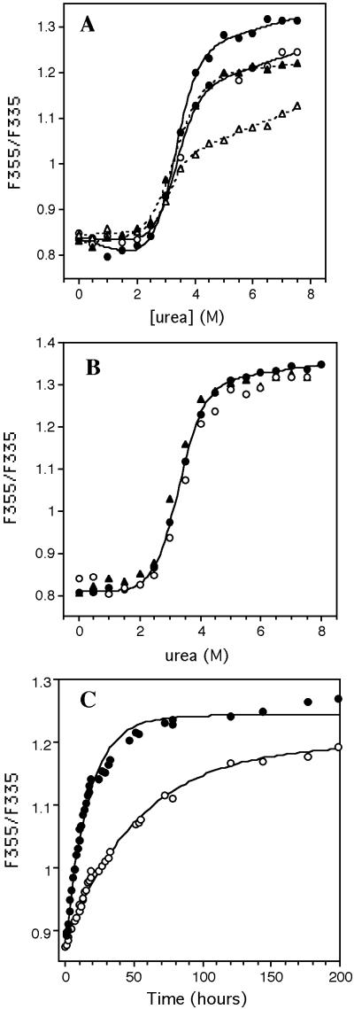
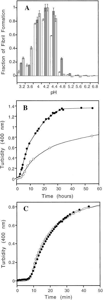
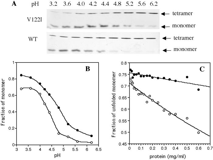
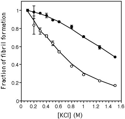
The transthyretin (TTR) amyloid diseases are of keen interest, because there are >80 mutations that cause, and a few mutations that suppress, disease. The V122I variant is the most common amyloidogenic mutation worldwide, producing familial amyloidotic cardiomyopathy primarily in individuals of African descent. The substitution shifts the tetramer-folded monomer equilibrium toward monomer (lowers tetramer stability) and lowers the kinetic barrier associated with rate-limiting tetramer dissociation (pH 7; relative to wild-type TTR) required for amyloid fibril formation. Fibril formation is also accelerated because the folded monomer resulting from the tetramer-folded monomer equilibrium rapidly undergoes partial denaturation and self-assembles into amyloid (in vitro) when subjected to a mild denaturation stress (e.g., pH 4.8). Incorporation of the V122I mutation into a folded monomeric variant of transthyretin reveals that this mutation does not destabilize the tertiary structure or alter the rate of amyloidogenesis relative to the wild-type monomer. The increase in the velocity of rate-limiting tetramer dissociation coupled with the lowered tetramer stability (increasing the mol fraction of folded monomer present at equilibrium) may explain why V122I confers an apparent absolute anatomic risk for cardiac amyloid deposition.


Figures

Figure 1
Figure 1
(A) Urea induced denaturation of V122I (circles) and WT (triangles) TTR, after incubation for 24 (open symbols) and 96 h (closed symbols), as detected by Trp fluorescence. Lines through the V122I (solid) and WT (dashed) data are smoothing curves to guide the eye. The lower value of WT plateau (at >3 M urea, 96 h) indicates this protein has not completely reached equilibrium. (B) Urea denaturation curves of monomeric V122I M-TTR (●) and WT M-TTR (▴) (20) (0.02 mg/ml 24 h incubation). That tetrameric V122I TTR (○) (96 h incubation) denaturation occurs via the monomer is demonstrated by the indistinguishable denaturation curves (● and ○, within error). The solid line through the V122I M-TTR data are fitted to a two-state model yielding a formula image value of 4.7 ± 0.2 kcal⋅mol−1 and m value of 1.4 ± 0.1 kcal⋅mol−1⋅M−1. (C) The rate of urea- (4.5 M) induced V122I (○) and WT (●) TTR tetramer dissociation detected by very fast linked tertiary structural changes. Solid lines are fitted to a first order single exponential function.
Figure 2
Figure 2
(A) Fibril formation of V122I TTR (0.2 mg/ml) monitored by turbidity (dark gray) and ThT binding (light gray) as a function of pH (37°C, 72 h). Fibril formation from WT TTR is shown by white bars, monitored by turbidity as a function of pH (37°C, 72 h). The relative amount of V122I fibril formation observed for the pH 4.2 sample was assigned to be unity (monitored by either ThT fluorescence or turbidity). The WT fibril yield is less than V122I at all pHs. (B) The rate of fibril formation at pH 4.2 (37°C) for V122I (●) and WT (○) TTR (0.2 mg/ml) ascertained by turbidity at 400 nm. (C) Kinetics of fibril formation from monomeric V122I M-TTR (●) and WT M-TTR (○) at 37°C (0.15 mg/ml, pH 4.4). The lines through the data points are smoothing curves to guide the eye.
Figure 3
Figure 3
(A) SDS/PAGE analysis of TTR quaternary structure as a function of pH. (Upper) V122I TTR; (Lower) WT TTR. (B) Mol fraction of monomer as a function of pH calculated by using densitometry from the gels shown in A. A smoothing curve applied to the V122I (●) and WT (○) data guides the eye. (C) Tetramer stability as a function of TTR concentration for V122I (●) and WT (○) in 4 M urea. Samples were incubated for 96 h at 25°C before analysis.
Figure 4
Figure 4
Inhibition of WT (○) and V122I (●) fibril formation as a function of Cl ion concentration. At 0.6 M Cl, 50% of WT fibrilization is inhibited, whereas V122I amyloidogenecity is reduced only by 10%.

Comment in

References

    1. Kelly J W. Curr Opin Struct Biol. 1996;6:11–17. - PubMed
    1. Buxbaum J N, Tagoe C E. Annu Rev Med. 2000;51:543–569. - PubMed
    1. Dobson C M. Trends Biochem Sci. 1999;24:329–332. - PubMed
    1. Fink A L. Folding Des. 1998;3:R9–R23. - PubMed
    1. Cohen A S, Connors L H. J Pathol. 1987;151:1–10. - PubMed

Publication types