. 2001 Dec 18;98(26):15020-5.
doi: 10.1073/pnas.011318098.

PDK1 regulates growth through Akt and S6K in Drosophila

Affiliations

PDK1 regulates growth through Akt and S6K in Drosophila

F Rintelen et al. Proc Natl Acad Sci U S A. .

Abstract

The insulin/insulin-like growth factor-1 signaling pathway promotes growth in invertebrates and vertebrates by increasing the levels of phosphatidylinositol 3,4,5-triphosphate through the activation of p110 phosphatidylinositol 3-kinase. Two key effectors of this pathway are the phosphoinositide-dependent protein kinase 1 (PDK1) and Akt/PKB. Although genetic analysis in Caenorhabditis elegans has implicated Akt as the only relevant PDK1 substrate, cell culture studies have suggested that PDK1 has additional targets. Here we show that, in Drosophila, dPDK1 controls cellular and organism growth by activating dAkt and S6 kinase, dS6K. Furthermore, dPDK1 genetically interacts with dRSK but not with dPKN, encoding two substrates of PDK1 in vitro. Thus, the results suggest that dPDK1 is required for dRSK but not dPKN activation and that it regulates insulin-mediated growth through two main effector branches, dAkt and dS6K.


Figures

Figure 1
Figure 1
Gain- and loss-of-function mutations in the dPDK1 locus. (a) Genomic structure of the dPDK1 locus. One of several reported transcripts (42) represented by the expressed sequence tag (EST) cDNA LD16509 is shown. Boxes represent exons. Dark boxes indicate the ORF; hatched and gray boxes represent the kinase and Pleckstrin-homology domains, respectively. EP insertions EP(3)0837, EP(3)3091, and EP(3)3644 are shown as triangles, and the direction of transcription from the UAS-controlled promoter is marked by arrows. EP(3)3644 inserted 7,081 and EP(3)0837 3,875 nt upstream of the putative start codon (Met). EP(3)3091 inserted 710 bp upstream of the 5′ end of exon 3. The three EMS-induced loss-of-function mutations and the activating mutation A467V are shown above exon 4. (b) Kinase domain alignment of Drosophila, C. elegans, and human PDK1. Dark and gray boxes indicate amino acid identity and similarity, respectively. Amino acid changes in the dPDK13–5 and dPDK1A467V mutants are shown above the dPDK1 sequence. Note that the amino acid substitutions in dPDK13 (G352S) and dPDK14 (P441L) are in highly conserved amino acid residues.
Figure 2
Figure 2
Simultaneous overexpression of dPDK1 and dAkt with GMR-Gal4 increases eye and cell size. (ad) Simultaneous overexpression of dPDK1 and dAkt in the developing third instar eye imaginal disk results in the formation of larger eyes. Scanning electron micrographs of adult eyes of the following genotypes: (a) OregonR, wild type; (b) y w; GMR-Gal4/+; EP(3)0837/+; (c) y w; GMR-Gal4 UAS-dAkt/+; TM2/+; (d) y w; GMR-Gal4 UAS-dAkt/+; TM2/EP(3)0837. Although the overexpression of dPDK1 or dAkt alone results only in a slight, but in the case of dPDK1, significant increase in eye size (b and c), simultaneous expression of dPDK1 and dAkt causes a substantial increase in eye size (d). The area of at least 29 ommatidia in 3–6 eyes was measured for each genotype. Because the size of ommatidia of the genotypes y w; GMR-Gal4 UAS-dAkt/+; TM2/+ and y w; GMR-Gal4 UAS-dAkt/+; TM2/EP(3)0837 is variable, only the values of the 30% largest ommatidia were included in the calculation. We used flies of the following genotype, y w; GMR-Gal4/UAS-lacZ, as a control. The means of these values are (normalized to a value of 100 ± SD): 100 ± 3 (control); 113 ± 3 (b); 108 ± 6 (c); 131 ± 10 (d). (eg) dPDK1 and dAkt act synergistically to increase cell size in a cell-autonomous manner. Tangential sections through adult eyes containing clones in which dPDK1 and/or dAkt were overexpressed: (e) y w hs-Flp/y w; GMR>FRT w+ STOP FRT>Gal4; EP(3)0837/+; (f) y w hs-Flp/y w; GMR>FRT w+ STOP FRT>Gal4/UAS-dAkt; (g) y w hs-Flp/y w; GMR>FRT w+ STOP FRT>Gal4/UAS-dAkt; EP(3)0837/+. Clones are marked by the lack of red pigment. No increase in cell size is observed by overexpressing dPDK1 or dAkt alone (e and f), but simultaneous overexpression of dPDK1 and dAkt slightly increases cell size (g). For quantification, the area of the R6 rhabdomere for 15 photoreceptors in clones overexpressing dPDK1 and/or dAkt (white arrowhead) were compared with the corresponding value for the sister control clone in the same section (yellow arrowhead): The values were normalized to 100 ± SD for the sister control clone and compared with the value in the overexpression clone: 100 ± 6 vs. 110 ± 7 (e); 100 ± 7 vs. 113 ± 7 (f); 100 ± 6 vs. 155 ± 10 (g). At the border of the clones, ommatidia composed of wild-type cells and cells overexpressing dPDK1 and/or dAkt are visible, indicating that the increase in cell size in g is cell-autonomous. (Bar = 100 μm.)
Figure 3
Figure 3
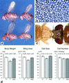
dPDK1 loss-of-function phenotypes. (a) Body size reduction of heteroallelic mutant flies. Males (Right) and females (Left) of the following genotypes are shown: y w; dPDK15/+ (Top); y w; dPDK14/dPDK15 (Bottom). (b) Tangential section through an eye containing a dPDK15/5 clone. Within the clone, all photoreceptor cells are reduced in size compared with wild-type photoreceptor cells. At the border of the clone, ommatidia composed of phenotypically wild-type and mutant cells (arrowhead) are visible, indicating that dPDK1 controls cell size autonomously. The genotype is as follows: y w ey-Flp/y w; dPDK15 FRT80B/FRT80B. (c) Selective removal of dPDK1 function from the eye imaginal disk results in a reduction of head and eye size. y w ey-Flp/y w; dPDK15 FRT80B/M(3)67c4 FRT80B (Left); OregonR, wild type (Right). (d) Quantification of body and organ size in dPDK1 heteroallelic mutant male flies compared with heterozygous and rescued flies, which overexpress a wild-type dPDK1 cDNA under the control of the ubiquitous arm-Gal4 driver. Values of y w; dPDK15/+ (+/−), y w; dPDK14/dPDK15 (−/−), and y w; arm-Gal4/UAS-dPDK1; dPDK14/dPDK15 flies (resc.) are shown. Values are the mean ± SD.
Figure 4
Figure 4
dPDK1 loss-of-function mutations suppress dInr and dPTEN mutant phenotypes. (a and b) The eye phenotype caused by overexpression of UAS-dInr with the GMR-Gal4 driver is dominantly suppressed by removing one copy of dPDK1, and the eye size is almost completely restored to wild-type size in a dPDK11/4 heteroallelic mutant background, although eye roughness is increased. The reason for this latter observation is unclear. (a) y w; GMR-Gal4 UAS-dInr/+; dPDK15/+ (Left); y w; GMR-Gal4 UAS-dInr/+; MKRS/+ (Right); (b) y w; GMR-Gal4 UAS-dInr/+; dPDK11/dPDK14 (Left); y w; GMR-Gal4 UAS-dInr/+; dPDK14/+ (Right). (c) The lethality caused by mutations in dPTEN is rescued in a dPDK1 heteroallelic mutant background: Some dPTEN, dPDK1 double-mutant flies survive to adulthood, although they display mutant phenotypes like an unproportionally reduced size of the abdomen and deformed leg structures. Similar phenotypes have been observed in partial loss-of-function mutations for dTOR (S. Oldham and E.H., unpublished work). Flies of the following genotypes are shown: y w dPTENdj189/dPTEN494; dPDK14/dPDK15 (Upper), OregonR, wild-type (Lower).
Figure 5
Figure 5
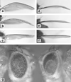
Genetic interaction of dPDK1 with the AGC kinases dAkt, dS6K, and dRSK. (ac) Mutations in dPDK1 suppress the ap-Gal4 UAS-dS6K bent-wing phenotype. (a) y w; ap-Gal4 UAS-dS6K/+; MKRS/+; (b) y w; ap-Gal4 UAS-dS6K/+; dPDK15/+; (c) y w; ap-Gal4 UAS-dS6K/+; dPDK14/dPDK15. (df) Null mutations in dS6K and dAkt dominantly suppress the ap-Gal4 UAS-dPDK1A467V bent-wing phenotype. (d) y w; ap-Gal4 UAS-dPDK1A467V/+; (e) y w; ap-Gal4 UAS-dPDK1A467V/+; dS6Kl-1/+; (f) y w; ap-Gal4 UAS-dPDK1A467V/+; dAkt1/+. (g) Mutations in dPDK1 suppress the rough eye phenotype caused by overexpression of UAS-dRSK under GMR-Gal4 control. y w; GMR-Gal4 UAS-dRSK/+; dPDK15/+ (Left); y w; GMR-Gal4 UAS-dRSK/+; dPDK14/dPDK15 (Right).

Similar articles

Cited by

References

    1. Alessi D R, James S R, Downes C P, Holmes A B, Gaffney P R, Reese C B, Cohen P. Curr Biol. 1997;7:261–269. - PubMed
    1. Alessi D R, Deak M, Casamayor A, Caudwell F B, Morrice N, Norman D G, Gaffney P, Reese C B, MacDougall C N, Harbison D, et al. Curr Biol. 1997;7:776–789. - PubMed
    1. Stokoe D, Stephens L R, Copeland T, Gaffney P R, Reese C B, Painter G F, Holmes A B, McCormick F, Hawkins P T. Science. 1997;277:567–570. - PubMed
    1. Stephens L, Anderson K, Stokoe D, Erdjument-Bromage H, Painter G F, Holmes A B, Gaffney P R, Reese C B, McCormick F, Tempst P, et al. Science. 1998;279:710–714. - PubMed
    1. Belham C, Wu S, Avruch J. Curr Biol. 1999;9:R93–R96. - PubMed

Publication types

MeSH terms

Substances