. 2001 Dec 18;98(26):15078-83.
doi: 10.1073/pnas.261569598.

The double par locus of virulence factor pB171: DNA segregation is correlated with oscillation of ParA

Affiliations

The double par locus of virulence factor pB171: DNA segregation is correlated with oscillation of ParA

G Ebersbach et al. Proc Natl Acad Sci U S A. .

Abstract

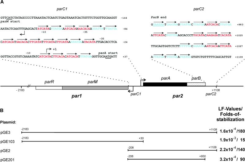
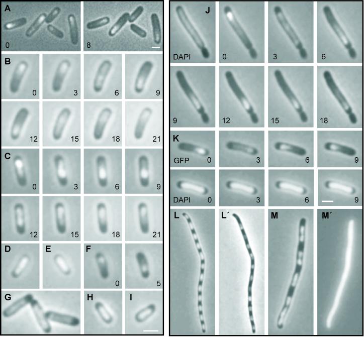
Prokaryotic plasmids and chromosomes encode partitioning (par) loci that segregate DNA to daughter cells before cell division. Recent database analyses showed that almost all known par loci encode an ATPase and a DNA-binding protein, and one or more cis-acting regions where the proteins act. All par-encoded ATPases belong to one of two protein superfamilies, Walker-type and actin-like ATPases. This property was recently used to divide par loci into Types I and II loci. We show here that the Escherichia coli virulence factor pB171 encodes a double par locus that consists of one Type I and one Type II locus. Separately, each locus stabilized a test-plasmid efficiently. Together, the two loci mediated even more efficient plasmid stabilization. The par loci have a unique genetic organization in that they share a common central region at which the two different DNA-binding proteins probably act. Interestingly, a fusion protein consisting of the Walker-type ParA ATPase and Gfp was functional and oscillated in nucleoid regions on a time scale of minutes. ParA-green fluorescent protein (Gfp) oscillation depended on both ParB and parC but was independent of minCDE. Point mutations in the Walker A box motif simultaneously abolished plasmid stabilization and ParA-Gfp oscillation. These observations raise the possibility that ParA oscillation is prerequisite for active plasmid segregation.


Figures

Figure 1
Figure 1
Overview of the par12 locus of pB171 (A) and of pB171-derived DNA fragments used in plasmid-stability tests (B). A shows the genetic organization of par1 and par2, parC1 and parC2. The gene encoding the actin-like ParM protein is shown as a gray box, parR as an open box, parA encoding the Walker-type ATPase as a black box, and parB is hatched. The DNA sequences of parC1 and parC2 are shown in blowups; arrows indicate direct repeats. Red, class I repeats; blue, class II repeats. Broken arrows pointing left and right indicate the transcription start points for the par1 and par2 operons, respectively. Numbering of base pairs according to the transcription start point of par2 (denoted +1). B shows extensions of PCR-derived DNA fragments inserted into the R1 test vector pRBJ200 and their effect on plasmid stability given as LF values measured as described by Gerdes et al. (40). Folds of stabilization relative to pRBJ200 (LF = 10−2 per cell per generation) of the respective plasmids are also shown. LF values are averages of five independent experiments for all four plasmids. LF values and their standard deviations: pGE3: (1.6 ± 0.55) × 10−4; pGE103: (1.9 ± 0.58) × 10−3; pGE2: (2.2 ± 0.87) × 10−4; pGE201: (3.2 ± 0.84) × 10−4.
Figure 2
Figure 2
Dynamic properties of functional ParA-Gfp in live cells. Combined phase contrast and fluorescent microscopy of ParA-Gfp-containing KG22 cells grown as described in Materials and Methods in the presence of 100 μM IPTG except in E and F (10 μM IPTG). Numbers are minutes in time-lapse experiments. (A) ParA-Gfp from pGE233 (par2 placparAgfp). Left and Right show the same cells; (B) a complete cycle of ParA-Gfp from pGE233 (par2 placparAgfp); (C) a complete oscillation cycle of ParA-Gfp from pGE236 (par2 ΔparA placparAgfp); (D) ParA-Gfp from pGE331 (parC1 placparAgfp); (E) ParA-Gfp from pGE230 (placparAgfp) in the presence of pGE223 (plachis6parB); (F) ParA-Gfp from pGE331 (parC1 placparAgfp) in the presence of pGE223 (plachis6parB); (G) ParA-Gfp from pGE230 (placparAgfp); (H and I) ParA-Gfp from pGE236G10V (par2 ΔparA placparA(G10V)∷gfp) and pGE236K14Q (par2 ΔparA placparA(K14Q)∷gfp), respectively; (J) ParA-Gfp from pGE236 (par2 ΔparA placparAgfp) in the minicell-producing strain M2141. DAPI staining was also included (Left). (K) DAPI and Gfp signals from cells of KG22 carrying pGE236 (par2 ΔparA placparAgfp); (L and M) DAPI staining of KG22/pGE230 (placparAgfp) and KG22/pGE30K (placlacZ′gfp) cells treated with cephalexin (10 μg/ml) and chloramphenicol (300 μg/ml). (L′ and M′) Gfp signal from the same cells. Omission of chloramphenicol yielded essentially similar although less clear patterns of nucleoid distribution. (Bar = 2 μm.)

Similar articles

Cited by

References

    1. Hiraga S. Annu Rev Genet. 2000;34:21–59. - PubMed
    1. Gordon G S, Wright A. Annu Rev Microbiol. 2000;54:681–708. - PubMed
    1. Møller-Jensen J, Jensen R B, Gerdes K. Trends Microbiol. 2000;8:313–320. - PubMed
    1. Gerdes K, Møller-Jensen J, Bugge J R. Mol Microbiol. 2000;37:455–466. - PubMed
    1. Watanabe E, Wachi M, Yamasaki M, Nagai K. Mol Gen Genet. 1992;234:346–352. - PubMed

Publication types