. 2001 Dec 18;98(26):15137-42.
doi: 10.1073/pnas.261570598.

T-bet is rapidly induced by interferon-gamma in lymphoid and myeloid cells

Affiliations

T-bet is rapidly induced by interferon-gamma in lymphoid and myeloid cells

A A Lighvani et al. Proc Natl Acad Sci U S A. .

Abstract

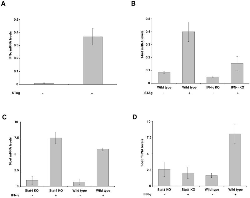
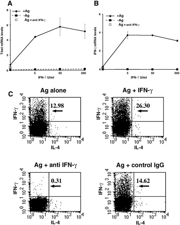
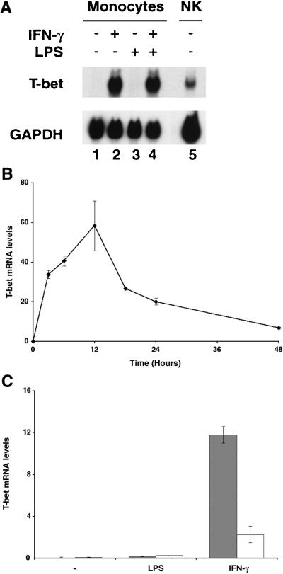
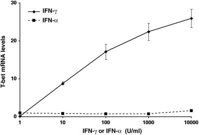
Differentiation of naive CD4(+) T cells into IFN-gamma-producing T helper 1 (T(H)1) cells is pivotal for protective immune responses against intracellular pathogens. T-bet, a recently discovered member of the T-box transcription factor family, has been reported to play a critical role in this process, promoting IFN-gamma production. Although terminal T(H)1 differentiation occurs over days, we now show that challenge of mice with a prototypical T(H)1-inducing stimulus, Toxoplasma gondii soluble extract, rapidly induced IFN-gamma and T-bet; T-bet induction was substantially lower in IFN-gamma-deficient mice. Naive T cells expressed little T-bet, but this transcription factor was induced markedly by the combination of IFN-gamma and cognate antigen. Human myeloid antigen-presenting cells showed T-bet induction after IFN-gamma stimulation alone, and this induction was antagonized by IL-4 and granulocyte/macrophage colony-stimulating factor. Although T-bet was induced rapidly and directly by IFN-gamma, it was not induced by IFN-alpha, lipopolysaccharide, or IL-1, indicating that this action of IFN-gamma was specific. Moreover, T-bet induction was dependent on Stat1 but not Stat4. These data argue for a model in which IFN-gamma gene regulation involves an autocrine loop, whereby the cytokine regulates a transcription factor that promotes its own production. These findings substantially alter the current view of T-bet in IFN-gamma regulation and promotion of cell-mediated immune responses.


Figures

Figure 1
Figure 1
IFN-γ signaling regulates T-bet expression. Murine splenocytes were collected from four wild-type or four IFN-γ−/− mice 6 h after T. gondii antigen challenge as well as from four untreated mice from each strain. IFN-γ (A) and T-bet (B) mRNA levels were measured by quantitative real-time reverse transcription–PCR (RT-PCR). (C) Splenocytes obtained from BALB/c/Stat4+/+ (wild type) and BALB/c/Stat4−/− (Stat4 KO) mice were treated with IFN-γ for 3 h. T-bet mRNA levels were assayed by quantitative real-time RT-PCR. (D) Splenocytes obtained from 129/SvEv/Stat1+/+ (wild type) and 129/SvEv/Stat1−/− (Stat1 KO) mice were treated with IFN-γ for 3 h in vitro. T-bet mRNA levels were assayed by quantitative real-time RT-PCR, using mRNA from Con-A-activated murine splenocytes for normalization. This level is arbitrarily designated as 1.0.
Figure 2
Figure 2
IFN-γ in conjunction with cognate antigen (Ag) induces T-bet and TH1 differentiation in naive CD4+ T cells. Naive CD4+ T cells were isolated from lymph nodes of 5C.C7 T cell receptor transgenic/Rag2-deficient mice, incubated with APCs and anti-IL-12 (10 μg/ml) in the presence (solid lines) or absence (dashed lines) of peptide antigen (100 μM) and various amounts of exogenous IFN-γ as indicated. Neutralizing anti-IFN-γ antibody (10 μg/ml) was added to selected cultures stimulated with peptide without exogenous IFN-γ (□). At 24 h, T-bet mRNA (A) and IFN-γ mRNA (B) levels were determined by quantitative RT-PCR. (C) In similar experiments, exogenous IFN-γ (5 units/ml) and anti-IFN-γ antibody blockade were analyzed for their effects on IFN-γ and IL-4 production, as detected by staining fixed, permeabilized cells for cytosolic cytokine content, at 72 h. Numbers in the upper right quadrant of each histogram indicate the percentage of IFN-γ-positive cells.
Figure 3
Figure 3
T-bet is induced rapidly in monocytes and DC by IFN-γ. Purified human monocytes were treated as indicated with IFN-γ and/or LPS, and T-bet mRNA levels were measured by Northern blotting (A Upper) and by real-time PCR (B). The experiment shown in A represents 3 h of stimulation; a glyceraldehyde-3-phosphate dehydrogenase (GAPDH) probe was used to confirm equal loading (A Lower). (C) Cultured human myeloid DC were either removed (shaded) or maintained (unshaded) in IL-4- and GM-CSF-containing medium before treatment with IFN-γ or LPS as indicated. T-bet levels were assayed by quantitative RT-PCR.
Figure 4
Figure 4
T-bet mRNA is induced specifically by IFN-γ but not IFN-α. Human monocytes were stimulated for 3 h with various concentrations of IFN-γ and IFN-α as indicated. T-bet mRNA levels were measured by real-time RT-PCR.

Similar articles

Cited by

References

    1. Abbas A K, Murphy K M, Sher A. Nature (London) 1996;383:787–793. - PubMed
    1. Murphy K M, Ouyang W, Farrar J D, Yang J, Ranganath S, Asnagli H, Afkarian M, Murphy T L. Annu Rev Immunol. 2000;18:451–494. - PubMed
    1. Bacon C M, Petricoin E F, III, Ortaldo J R, Rees R C, Larner A C, Johnston J A, O'Shea J J. Proc Natl Acad Sci USA. 1995;92:7307–7311. - PMC - PubMed
    1. Jacobson N G, Szabo S J, Guler M L, Gorham J D, Murphy K M. Adv Exp Med Biol. 1996;409:61–73. - PubMed
    1. Kaplan M H, Sun Y L, Hoey T, Grusby M J. Nature (London) 1996;382:174–177. - PubMed

Publication types

MeSH terms