. 2001 Dec 18;98(26):15221-6.
doi: 10.1073/pnas.261572998.

Powerful and prolonged protection of human retinal pigment epithelial cells, keratinocytes, and mouse leukemia cells against oxidative damage: the indirect antioxidant effects of sulforaphane

Affiliations

Powerful and prolonged protection of human retinal pigment epithelial cells, keratinocytes, and mouse leukemia cells against oxidative damage: the indirect antioxidant effects of sulforaphane

X Gao et al. Proc Natl Acad Sci U S A. .

Abstract

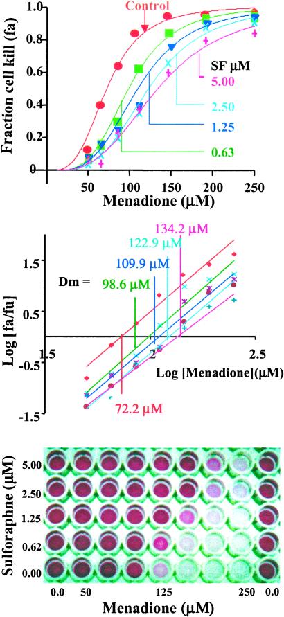
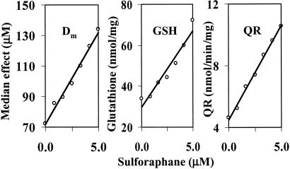
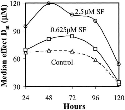
Mammalian cells are equipped with elaborate systems for protection against the toxicity of reactive oxygen and nitrogen species and electrophiles that are constant dangers to the integrity of their DNA. Phase 2 enzymes (e.g., glutathione transferases, NAD(P)H:quinone reductase) and glutathione synthesis are widely recognized as playing major protective roles against electrophilic carcinogens, but their antioxidant functions have attracted far less attention. The cytotoxicities of four oxidative stressors (menadione, tert-butyl hydroperoxide, 4-hydroxynonenal, and peroxynitrite) for human adult retinal pigment epithelial cells (ARPE-19) were quantified by measuring the concentration dependence of cell death and were expressed as the median effect dose (D(m)) for each oxidant. After treatment of ARPE-19 cells for 24 h with 0-5 microM concentrations of sulforaphane (the powerful Phase 2 enzyme inducer isolated from broccoli), the toxicities of the oxidants were markedly reduced as shown by 1.5- to 3-fold increases in D(m) values. The magnitude of protection was a function of the nature of the oxidants and the concentrations of both the oxidants and sulforaphane. Protection was prolonged and persisted for several days after removal of sulforaphane before returning to control levels. The sulforaphane-dependent increases in specific activities of cytosolic quinone reductase and the glutathione levels were highly significantly correlated with the degree of protection as measured by D(m) values. Antioxidant protection was also demonstrated for human HaCaT keratinocytes and L1210 murine leukemia cells. It is therefore highly likely that the multifaceted and prolonged antioxidant protection provided by sulforaphane is a general phenomenon that is mediated through induction of the Phase 2 enzyme response.


Figures

Figure 1
Figure 1
Protection of adult human retinal pigment epithelial (ARPE-19) cells against the toxicity of menadione (0–250 μM) by induction of Phase 2 enzymes by sulforaphane (SF) (0–5 μM). (Upper) Fractional killing of cells (fa) as a function of menadione concentration at a series of sulforaphane concentrations. (Center) Analysis of the data by the median effect plot. The median effect concentrations (Dm) at the sulforaphane concentrations above. (Lower) Photograph of a typical 96-well microtiter plate showing the protective effect of sulforaphane against the cytotoxicity of menadione for human ARPE-19 cells. The intensity of purple color is the reduced MTT formazan for a measure of cell viability.
Figure 2
Figure 2
Comparison of the effects of treatment of human ARPE-19 cells with a series of concentrations of sulforaphane (0–5 μM) for 24 h on the toxicity of exposure for 2 h to menadione. (Left) Cytotoxicity expressed as the median effect concentration (Dm). (Center) Glutathione concentrations expressed as nanomoles per milligram of cytosolic protein. (Right) Quinone reductase specific activity, expressed as nanomoles per minute per milligram of cytosolic protein. The multivariate regression correlations between sulforaphane concentrations and the other three variables all had P values of <0.01.
Figure 3
Figure 3
Prolonged protection of ARPE-19 cells against menadione toxicity by sulforaphane (SF) expressed as median effect concentrations (Dm, μM). The menadione toxicity was determined immediately after induction (time = 0), and 24, 48, 96, and 120 h later. Note that protection continued to rise for 24–48 h, and then declined during the next 48 h.
Figure 4
Figure 4
Persistent induction of quinone reductase (QR), glucose-6-phosphate dehydrogenase (G6PD), glutathione reductase (GR) (nanomoles per minute per milligram of cytosolic protein) and elevation of GSH levels (expressed as nanomoles per milligram of cytosolic protein) in human ARPE-19 cells after exposure to sulforaphane [0 (▴), 0.625 (□), and 2.5 (●) μM] for 24 h.
Figure 5
Figure 5
Protection of human ARPE-19 cells against the toxicity of menadione, tert-butyl hydroperoxide (0.5, 0.75, 1 mM for 16 h), 4-hydroxynonenal (6.25, 12.5, 25 μM for 4 h), and peroxynitrite (1, 2, 4 mM for 2 h) as a function of prior exposure for 24 h to 0–2.5 μM sulforaphane. The bar graphs show that cell viability is a function of both the concentrations of the oxidant and of the sulforaphane inducer. The front, center, and rear series of bars refer to the highest, middle, and lowest concentration of oxidants, respectively.
Figure 6
Figure 6
Protection of human keratinocytes (HaCaT) against oxidative cytotoxicity of tert-butyl hydroperoxide (0.313, 0.625, 1 mM for 8 h) (Left) and mouse leukemia (L1210) cells against oxidative cytotoxicity of menadione (15.6, 31.3, 62.5 μM for 2 h) (Right) by treatment with sulforaphane (0–2.5 μM for 24 h). The Dm and m values obtained from the median effect plots are shown in Table 1. The front, center, and rear series of bars refer to the highest, middle, and lowest concentration of oxidants, respectively.

Similar articles

Cited by

References

    1. Halliwell B, Gutteridge J M C. Free Radicals in Biology and Medicine. New York: Oxford Univ. Press; 1999. pp. 1–36.
    1. Pokorny J, Yanishlieva M, Gordon M. Antioxidants in Food: Practical Applications. Cambridge, U.K.: Woodhead Publishing, Ltd.; 2001.
    1. Fahey J W, Zhang Y, Talalay P. Proc Natl Acad Sci USA. 1997;94:10367–10372. - PMC - PubMed
    1. Kensler T W. Environ Health Perspect. 1997;105:965–970. - PMC - PubMed
    1. Talalay P, Fahey J W, Holtzclaw W D, Prestera T, Zhang Y. Toxicol Lett. 1995;82/83:173–179. - PubMed

Publication types