. 2001 Dec 18;98(26):15276-81.
doi: 10.1073/pnas.261574998.

Neisseria gonorrhoeae PilV, a type IV pilus-associated protein essential to human epithelial cell adherence

Affiliations

Neisseria gonorrhoeae PilV, a type IV pilus-associated protein essential to human epithelial cell adherence

H C Winther-Larsen et al. Proc Natl Acad Sci U S A. .

Abstract

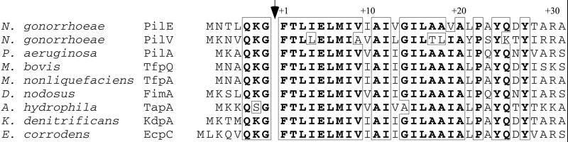
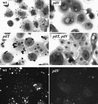
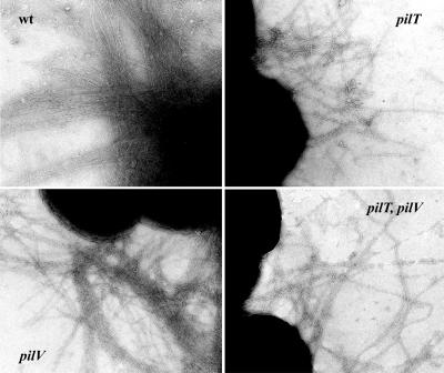
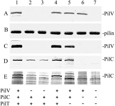
Type IV pili (Tfp) of Neisseria gonorrhoeae, the Gram-negative etiologic agent of gonorrhea, facilitate colonization of the human host. Tfp are assumed to play a key role in the initial adherence to human epithelial cells by virtue of the associated adhesin protein PilC. To examine the structural and functional basis for adherence in more detail, we identified potential genes encoding polypeptides sharing structural similarities to PilE (the Tfp subunit) within the N. gonorrhoeae genome sequence database. We show here that a fiber subunit-like protein, termed PilV, is essential to organelle-associated adherence but dispensable for Tfp biogenesis and other pilus-related phenotypes, including autoagglutination, competence for natural transformation, and twitching motility. The adherence defect in pilV mutants cannot be attributed to reduced levels of piliation, defects in fiber anchoring to the bacterial cell surface, or to unstable pilus expression related to organelle retraction. PilV is expressed at low levels relative to PilE and copurifies with Tfp fibers in a PilC-dependent fashion. Purified Tfp from pilV mutants contain PilC adhesin at reduced levels. Taken together, these data support a model in which PilV functions in adherence by promoting the functional display of PilC in the context of the pilus fiber.


Figures

Figure 1
Figure 1
N-terminal aligment of PilV with Tfp prepilins. The arrow indicates the prepilin peptidase processing site of the prepilin proteins and predicted processing site of PilV. Identical residues are boxed and in bold.
Figure 2
Figure 2
Adherence of gonococcal strains and Tfp to human corneal epithelial cells. (Top and Middle) Adherent cells are stained with crystal violet. wt (N400; recA6(tetM)); pilV (GV2; pilV∷kan); pilT (MW4; pilTind(tetM)); and PilT, pilV (GV4; pilV∷kan, pilTind). (Bottom) Binding of Tfp to human corneal epithelial cells assesed by indirect immunofluorescence. wt (N400; recA6(tetM)); and pilV (GV2; pilV∷kan). All panels are shown at the same level of magnification.
Figure 3
Figure 3
Piliation of gonococcal strains analyzed by transmission electron microscopy. wt (N400, recA6(tetM)); pilV (GV2, pilV∷kan); pilT (MW4, pilTind (tetM)); and pilT, pilV (GV4, pilV∷kan, pilTind). Micrographs are taken at a magnification of ×90,000.
Figure 4
Figure 4
Quantitative analysis of PilV and PilC in whole cells and purified pili from gonococcal strains. Lanes 1, N401 (wt); 2, GV1 (pilVG-1 fs); 3, GV2 (pilV∷kan); 4, GV3 (pilV∷kan, iga∷pilV); 5, MW4 (pilTind); 6, MW7 (pilTind, pilC1∷erm, pilC2∷cat); and 7, GV7 (pilVG-1 fs,, pilTind, pilC1∷erm, pilC2∷cat). (A) Immunoblotting of whole cell lysates by using rabbit antibodies specific for PilV. (B) Coomassie-stained SDS/PAGE gel showing the relative amounts of PilE in purified pili. (C) Immunoblotting of purified pili by using rabbit antibodies specific for PilV. (D) Immunoblotting of purified pili by using rabbit antibodies specific for PilC. (E) Detection of PilC by colloidal silver staining of purified pilus proteins transferred to a poly(vinylidene difluoride) membrane. The amounts of samples loaded in A were equalized based on the total protein concentration of whole cells. The amounts of purified pili loaded in BE, lanes 1 through 4, were standardized based on the weight of whole cells from which the preparations were made. The comparable intensities of the Coomassie-stained PilE band in B (reflecting its relative yield) provide a quantitative readout of piliation levels (19). The amounts of pili loaded in lanes 5–7 were adjusted to be equivalent to those used in lanes 1–4.

Similar articles

Cited by

References

    1. Soto G E, Hultgren S J. J Bacteriol. 1999;181:1059–1071. - PMC - PubMed
    1. Sauer F G, Barnhart M, Choudhury D, Knight S D, Waksman G, Hultgren S J. Curr Opin Struct Biol. 2000;10:548–556. - PubMed
    1. Strom M S, Lory S. Annu Rev Microbiol. 1993;47:565–596. - PubMed
    1. Wolfgang M, van Putten J P, Hayes S F, Dorward D, Koomey M. EMBO J. 2000;19:6408–6418. - PMC - PubMed
    1. Heckels J E. Clin Microbiol Rev. 1989;2:S66–S73. - PMC - PubMed

Publication types

MeSH terms

Associated data

LinkOut - more resources