. 2001 Dec 18;98(26):15330-5.
doi: 10.1073/pnas.261273898.

Thalamic bursting in rats during different awake behavioral states

Affiliations

Thalamic bursting in rats during different awake behavioral states

E E Fanselow et al. Proc Natl Acad Sci U S A. .

Abstract

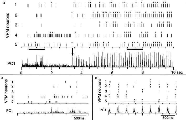
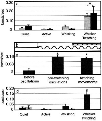
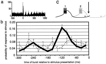
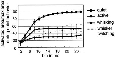
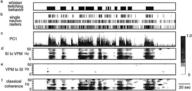
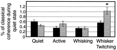
Thalamic neurons have two firing modes: tonic and bursting. It was originally suggested that bursting occurs only during states such as slow-wave sleep, when little or no information is relayed by the thalamus. However, bursting occurs during wakefulness in the visual and somatosensory thalamus, and could theoretically influence sensory processing. Here we used chronically implanted electrodes to record from the ventroposterior medial thalamic nucleus (VPM) and primary somatosensory cortex (SI) of awake, freely moving rats during different behaviors. These behaviors included quiet immobility, exploratory whisking (large-amplitude whisker movements), and whisker twitching (small-amplitude, 7- to 12-Hz whisker movements). We demonstrated that thalamic bursting appeared during the oscillatory activity occurring before whisker twitching movements, and continued throughout the whisker twitching. Further, thalamic bursting occurred during whisker twitching substantially more often than during the other behaviors, and a neuron was most likely to respond to a stimulus if a burst occurred approximately 120 ms before the stimulation. In addition, the amount of cortical area activated was similar to that during whisking. However, when SI was inactivated by muscimol infusion, whisker twitching was never observed. Finally, we used a statistical technique called partial directed coherence to identify the direction of influence of neural activity between VPM and SI, and observed that there was more directional coherence from SI to VPM during whisker twitching than during the other behaviors. Based on these findings, we propose that during whisker twitching, a descending signal from SI triggers thalamic bursting that primes the thalamocortical loop for enhanced signal detection during the whisker twitching behavior.


Figures

Figure 1
Figure 1
Neural activity in VPM during quiet immobility and whisker twitching behavior. (a) During quiet immobility (before arrow in PC1), neuronal firing was not periodic and did not frequently involve bursting. However, after the oscillations began, the firing was very phasic and tended to occur in bursts. Note that not every neuron fired on every cycle. (b and c) Enlargements of activity in the left and right dark bars in a, respectively. Asterisks indicate activity that qualified as bursts according to the criteria described in Methods; note that individual spikes cannot be resolved at this time scale.
Figure 2
Figure 2
Burst firing in VPM and SI. (a) During the whisker twitching behavior there was significantly more bursting activity in both VPM and SI than during the other three behavioral states. Gray bars are for VPM and black bars are for SI. Asterisk and bracket indicate that the VPM and SI values are both different from values during the other behaviors. (b and c) The amount of burst activity increased relative to baseline during both the pre-twitching oscillations (after arrow in b) and after the whisker twitching movements began (hatched bar in b). The burst rate during the pre-twitching oscillations was not significantly different from that after the whisker twitching movements began. Asterisks in c indicate values significantly different from the “before oscillations” condition. (d) After inactivation of SI cortex, bursting activity in VPM was still higher during whisker twitching than during any of the other behaviors. Black bars show premuscimol and gray bars show postmuscimol values. Asterisk indicates value is significantly different from all others.
Figure 3
Figure 3
Resetting of oscillation phase and probability of a response to infraorbital nerve stimulation after a burst. (a) Histogram showing that the phase of oscillations during whisker twitching is reset with the presentation of a stimulus (vertical broken line). (b) The probability of a response to the stimulus depended on when the stimulus was presented relative to the timing of the previous burst. (c) This schematic diagram illustrates this point by showing a “burst” of action potentials and the period after the burst when the cell is unable to fire, relative to when the stimulus occurs (arrow/0 ms on graph in b). Asterisks show approximately where the IT current becomes de-inactivated and the cell is ready to fire. If the burst labeled 1 occurs, the neuron is likely to respond when the stimulus occurs, whereas if burst 2 occurs instead, the neuron is much less likely to fire in response to the stimulus.
Figure 4
Figure 4
Cumulative sum of activated cortical area. This graph shows the total amount of cortical area activated in the first 28 ms after stimulation of the infraorbital nerve. Values are presented as a percent of cumulative area activated during the quiet behavior.
Figure 5
Figure 5
PDC. The traces in this figure are from 250 s of a recording that includes periods of whisker twitching behavior. (a) Bars indicate times when the rat was engaged in the whisker twitching behavior. (b) Raster plots showing spike times for three single-neuron recordings. (c) PC1 of activity in VPM. (d) PDC from SI to VPM. (e) PDC from VPM to SI. (f) Classical coherence between VPM and SI. Gray scale bar indicates fraction of total power at each frequency for a given source (VPM or SI).
Figure 6
Figure 6
PDC during different behavioral states. During the whisker twitching state there was a significantly larger amount of directed coherence from SI to VPM than from VPM to SI (∗). In addition, the amount of directed coherence between SI and VPM during whisker twitching was larger than during any other behavioral state. Black bars show directed coherence from VPM to SI, and gray bars show directed coherence from SI to VPM. Values were normalized to the amount of classical coherence for each measure.

Similar articles

Cited by

References

    1. McCarley R W, Benoit O, Barrionuevo G. J Neurophysiol. 1983;50:798–818. - PubMed
    1. McCormick D A. J Neurosci. 1992;12:278–289. - PMC - PubMed
    1. Maffei L, Moruzzi G, Rizzolatti G. Arch Ital Biol. 1965;103:596–608. - PubMed
    1. Sherman S M, Guillery R W. J Neurophysiol. 1996;76:1367–1395. - PubMed
    1. Sherman S M. Trends Neurosci. 2001;24:122–126. - PubMed

Publication types