

Primary antiphospholipid antibody syndrome: neuroradiologic findings in 11 patients
- PMID: 11752922
- PMCID: PMC2718138
- DOI: 10.3348/kjr.2000.1.1.5
Primary antiphospholipid antibody syndrome: neuroradiologic findings in 11 patients
Abstract
Objective: To describe the neuroradiologic findings of primary antiphospholipid antibody syndrome (PAPS).
Materials and methods: During a recent two-year period, abnormally elevated antiphospholipid antibodies were detected in a total of 751 patients. In any cases in which risk factors for stroke were detected-hypertension, diabetes mellitus, hyperlipidemia, smoking, and the presence of SLE or other connective tissue diseases-PAPS was not diagnosed. Neuroradiologic studies were performed in 11 of 32 patients with PAPS. We retrospectively reviewed brain CT (n = 7), MR (n = 8), and cerebral angiography (n = 8) in 11 patients with special attention to the presence of brain parenchymal lesions and cerebral arterial or venous abnormalities.
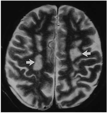
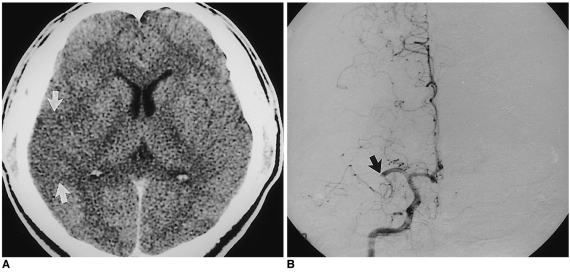
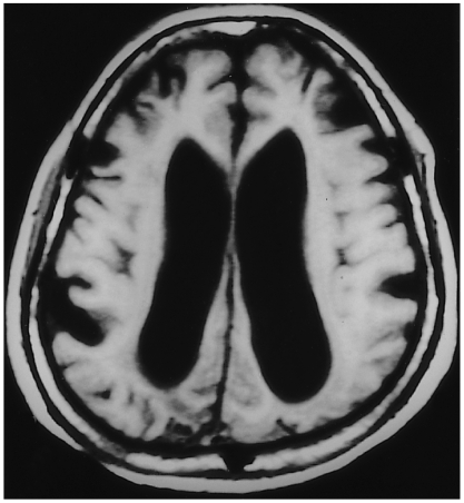
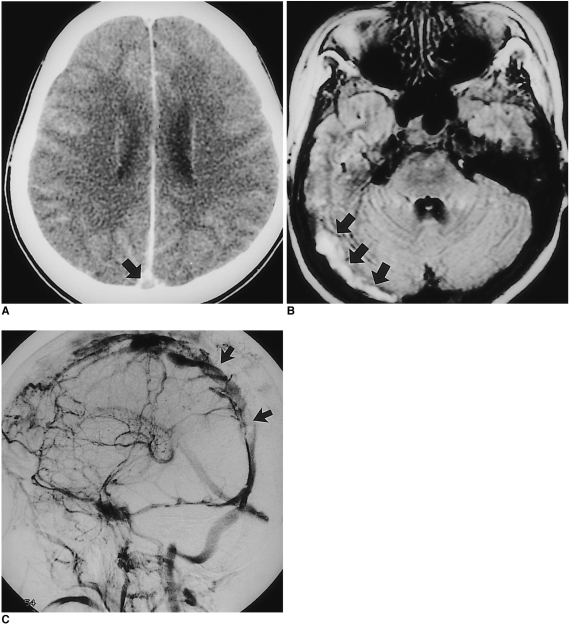
Results: CT or MR findings of PAPS included nonspecific multiple hyper-intensity foci in deep white matter on T2-weighted images (5/11), a large infarct in the territory of the middle cerebral artery (4/11), diffuse cortical atrophy (2/11), focal hemorrhage (2/11), and dural sinus thrombosis (1/11). Angiographic findings were normal (5/8) or reflected either occlusion of a large cerebral artery (2/8) or dural sinus thrombosis (1/8).
Conclusion: Neuroradiologic findings of PAPS are nonspecific but in young or middle-aged adults who show the above mentioned CT or MR findings, and in whom risk factors for stroke are not present, the condition should be suspected.
Figures
References
-
- Harris EN, Gharavi AE, Hughes GRV. Antiphospholipid antibodies. Clin Rheum Dis. 1985;11:591–609. - PubMed
-
- Harris EN, Baguley E, Asherson RA, et al. Clinical and serological features of the "antiphospholipid syndrome". Br J Rheumatol. 1987;26(S):19.
-
- Hughson MD, Mccarty GA, Brumback RA. Spectrum of vascular pathology affecting patients with the antiphospholipid syndrome. Hum Pathol. 1995;26:716–724. - PubMed
-
- Asherson RA, Khamashta MA, Ordi-Ros J, et al. The "primary" antiphospholiupid syndrome: major clinical and serological features. Medicine. 1989;68:366–374. - PubMed
-
- Gharavi AE, Wilson WA. The syndrome of thrombosis, thrombocytopenia, and recurrent spontaneous abortions associated with antiphospholipid antibodies: Hughes syndrome. Lupus. 1996;5:343–344. - PubMed
MeSH terms
LinkOut - more resources
Full Text Sources
