. 2000 Oct-Dec;1(4):185-90.
doi: 10.3348/kjr.2000.1.4.185.

Hepatic cavernous hemangioma in cirrhotic liver: imaging findings

Affiliations

Hepatic cavernous hemangioma in cirrhotic liver: imaging findings

J S Yu et al. Korean J Radiol. 2000 Oct-Dec.

Abstract

Objective: To document the imaging findings of hepatic cavernous hemangioma detected in cirrhotic liver.

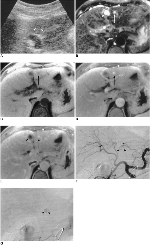
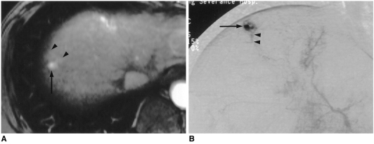
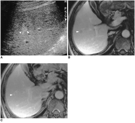
Materials and methods: The imaging findings of 14 hepatic cavernous hemangiomas in ten patients with liver cirrhosis were retrospectively analyzed. A diagnosis of hepatic cavernous hemangioma was based on the findings of two or more of the following imaging studies: MR, including contrast-enhanced dynamic imaging (n = 10), dynamic CT (n = 4), hepatic arteriography (n = 9), and US (n = 10).

Results: The mean size of the 14 hepatic hemangiomas was 0.9 (range, 0.5-1.5) cm in the longest dimension. In 11 of these (79%), contrast-enhanced dynamic CT and MR imaging showed rapid contrast enhancement of the entire lesion during the early phase, and hepatic arteriography revealed globular enhancement and rapid filling-in. On contrast-enhanced MR images, three lesions (21%) showed partial enhancement until the 5-min delayed phases. US indicated that while three slowly enhancing lesions were homogeneously hyperechoic, 9 (82%) of 11 showing rapid enhancement were not delineated.

Conclusion: The majority of hepatic cavernous hemangiomas detected in cirrhotic liver are small in size, and in many, hepatic arteriography and/or contrast-enhanced dynamic CT and MR imaging demonstrates rapid enhancement. US, however, fails to distinguish a lesion of this kind from its cirrhotic background.


Figures

Fig. 1
Fig. 1
A 62-year-old woman with hypoechoic, rapidly enhancing small hemangioma detected in a cirrhotic liver caused by chronic B-viral hepatitis. A. US depicts a 0.8-cm hypoechoic tumor (arrowheads) in the left lobe of the liver. B. T2-weighted turbo spin-echo MR image (TR/TE, 4060/138) shows a tumor (arrow) with bright signal intensity. Another high signal intensity tumor is seen (arrowhead), and this is a simple cyst. C. Unenhanced T1-weighted spoiled gradient-echo MR image with fat suppression (160.8/4.1) shows a low signal intensity tumor (arrow). D. Arterial dominant phase spoiled gradient-echo image (160.8/4.1) reveals strong enhancement of the tumor (arrow) seen in E. E. Delayed phase spoiled gradient-echo image (160.8/4.1) obtained 5 min after the start of contrast injection demonstrates persistent contrast enhancement of the tumor (arrow). Simple cyst (arrowhead) is not enhanced after contrast injection. F. Arterial phase of hepatic arteriography shows small C-shaped contrast puddling (arrowheads). G. Capillary phase of hepatic arteriography depicts more diffuse contrast accumulation in the lesion (arrowheads) seen in F.
Fig. 2
Fig. 2
A 45-year-old man with small rapidly enhancing hemangioma in a cirrhotic liver resulting from chronic B-viral hepatitis. A. Arterial dominant phase contrast-enhanced spoiled gradient-echo MR image (140/2.7) shows strong enhancement of a 0.9-cm tumor (arrow) with wedge-shaped temporal peritumoral enhancement (arrowheads). On unenhanced T1-and T2-weighted, and delayed contrast-enhanced MR images (not shown), the area of wedge-shaped enhancement could not be distinguished from surrounding hepatic parenchyma. B. Capillary phase of hepatic arteriography shows near complete filling-in of the tumor by contrast agent (arrow), with opacification of a small, proximal portal vein branch (arrowheads), suggesting a transtumoral shunt or drainage of the hyperdynamic tumor by this branch.
Fig. 3
Fig. 3
A 52-year-old man with hyperechoic, slowly-enhancing small hemangima detected in a cirrhotic liver caused by chronic B-viral hepatitis. A. US shows 1.1-cm hyperechoic tumor (arrowheads) in the right lobe of the liver. B. Parenchymal phase contrast-enhanced spoiled gradient-echo MR image (140/2.7) demonstrates minimal tumoral enhancement (arrowhead). C. Delayed phase contrast-enhanced spoiled gradient-echo MR image (140/2.7) obtained 5 mins after the administration of contrast agent depicts more diffuse, but still partial, contrast enhancement of the tumor (arrowhead) seen in B. Hepatic arteriogram (not shown) demonstrated no tumoral contrast enhancement.

References

    1. Itai Y, Ohnishi S, Ohtomo K, Kokubo T, Imawari M, Atomi Y. Hepatic cavernous hemangioma in patients at high risk for liver cancer. Acta Radiol. 1987;28:697–701. - PubMed
    1. Hanafusa K, Ohashi I, Himeno Y, Suzuki S, Shibuya H. Hepatic hemangioma: findings with two-phase CT. Radiology. 1995;196:465–469. - PubMed
    1. Takayasu K, Moriyama N, Shima Y, et al. Atypical radiographic findings in hepatic cavernous hemangioma: correlation with histologic features. AJR. 1986;146:1149–1153. - PubMed
    1. Moody AR, Wilson SR. Atypical hepatic hemangioma: a suggestive sonographic morphology. Radiology. 1993;188:413–417. - PubMed
    1. Freeny PC, Vimont TR, Barnett DC. Cavernous hemangioma of the liver: ultrasonography, arteriography, and computed tomography. Radiology. 1979;132:143–148. - PubMed