

Recurrent uterine cervical carcinoma: spectrum of imaging findings
- PMID: 11752955
- PMCID: PMC2718201
- DOI: 10.3348/kjr.2000.1.4.198
Recurrent uterine cervical carcinoma: spectrum of imaging findings
Abstract
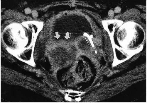
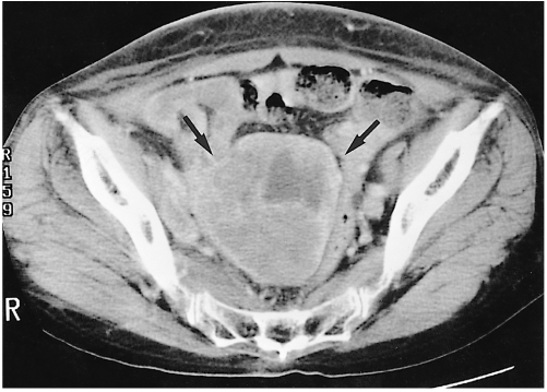
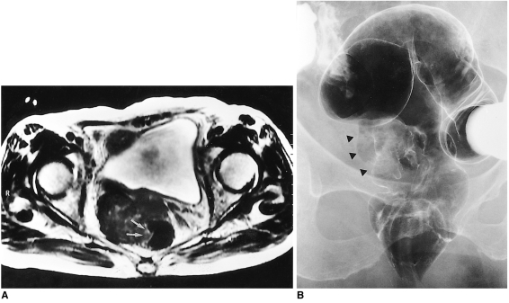
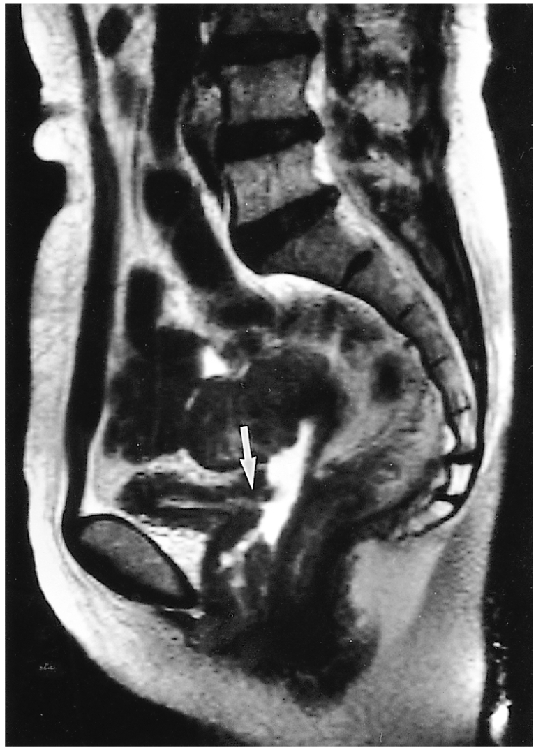
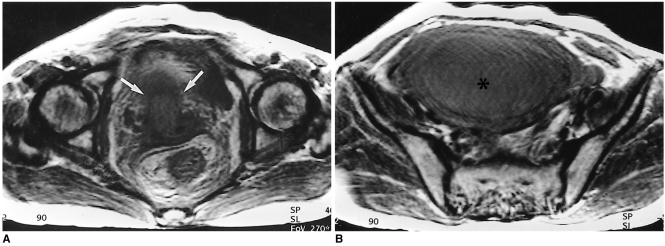
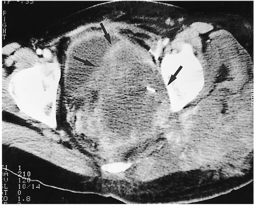
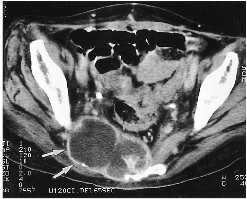
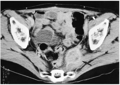
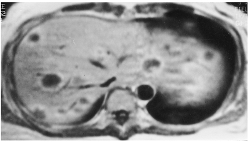
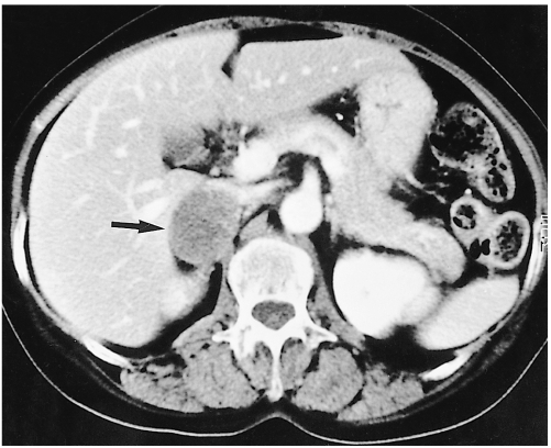
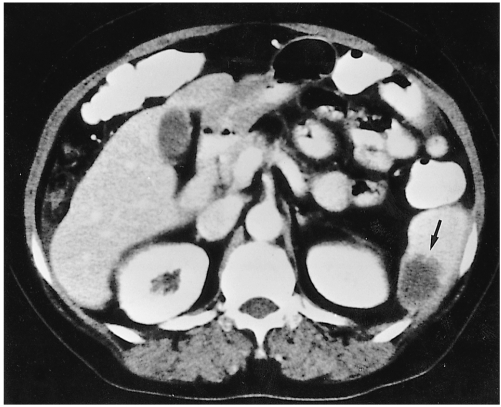
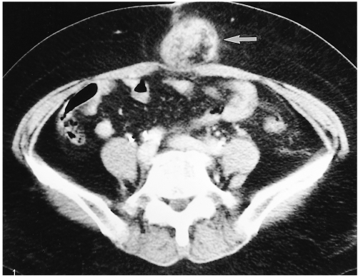
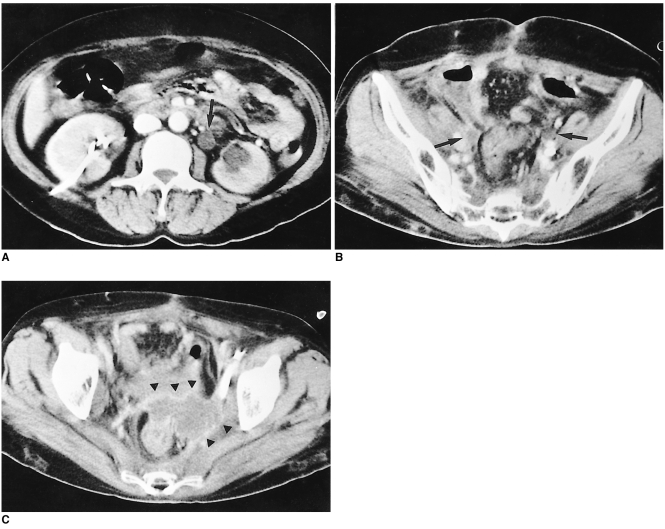
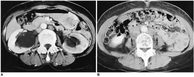
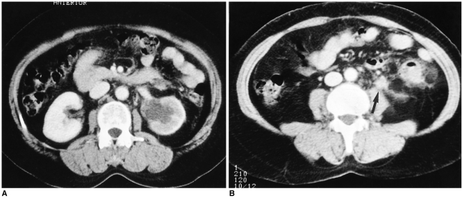
Uterine cervical carcinoma is one of the most common malignant tumors occurring in females. After primary treatment, patients are usually followed up with CT or MRI and the findings of these modalities may be the first sign of recurrent disease. Because earlier additional treatment by chemotherapy or radiation therapy may improve the prognosis, the early detection of recurrent cervical carcinoma is clinically important. In this article, we review the CT and MR imaging findings of recurrent uterine cervical carcinoma, and assign them to one of four groups: a) recurrence at the primary site, involving the intrapelvic organs, b) extension to the pelvic side-wall, c) metastases to pelvic and extrapelvic lymph nodes, or d) metastases to distant organs. A further contribution of CT and MR imaging is the detection of hydronephrosis due to ureteral obstruction. The cases in each group are illustrated and discussed, and since an awareness of the spectrum of imaging findings of recurrent cervical carcinoma is likely to lead to its early detection, radiologists should be familiar with the information presented.
Figures
References
-
- Walsh JW. Computed tomography of the pelvis. Churchill Livingstone: New York; 1985. pp. 195–198.
-
- Carlson V, Delclos L, Fletcher GH. Distant metastases in squamous-cell carcinoma of the uterine cervix. Radiology. 1967;88:961–966. - PubMed
-
- Walsh JW, Amendola MA, Hall DJ, Tisnado J, Goplerud DR. Recurrent carcinoma of the pelvis: CT diagnosis. AJR. 1981;136:117–122. - PubMed
-
- Fulcher AS, O'Sullivan SG, Segreti EM, Kavanagh BD. Recurrent cervical carcinoma: Typical and atypical manifestations. RadioGraphics. 1999;19:s103–s116. - PubMed
-
- Kim JE, Park HA, Kim KH, Lim D, Chin SY. Patterns of recurrent cervical carcinoma on CT. J Korean Radiol Soc. 1988;24:1130–1134.
