

Unenhanced spiral CT in acute ureteral colic: a replacement for excretory urography?
- PMID: 11752964
- PMCID: PMC2718090
- DOI: 10.3348/kjr.2001.2.1.14
Unenhanced spiral CT in acute ureteral colic: a replacement for excretory urography?
Abstract
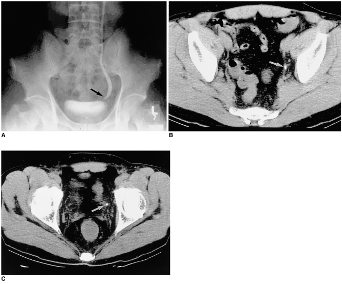
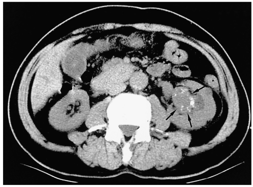
Objective: To compare the usefulness of unenhanced spiral CT (UCT) with that of excretory urography (EU) in patients with acute flank pain.
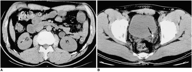
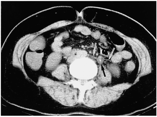
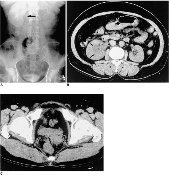
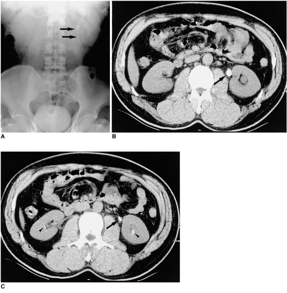
Materials and methods: Thirty patients presenting with acute flank pain underwent both UCT and EU. Both techniques were used to determine the presence, size, and location of urinary stone, and the presence or absence of secondary signs was also evaluated. The existence of ureteral stone was confirmed by its removal or spontaneous passage during follow-up. The absence of a stone was determined on the basis of the clinical and radiological evidence.
Results: Twenty-one of the 30 patients had one or more ureteral stones and nine had no stone. CT depicted 22 of 23 calculi in the 21 patients with a stone, and no calculus in all nine without a stone. The sensitivity and specificity of UCT were 96% and 100%, respectively. EU disclosed 14 calculi in the 21 patients with a stone and no calculus in eight of the nine without a stone. UCT and EU demonstrated secondary signs of ureterolithiasis in 15 and 17 patients, respectively.
Conclusion: For the evaluation of patients with acute flank pain, UCT is an excellent modality with high sensitivity and specificity. In near future it may replace EU.
Figures
References
-
- Sierakowski R, Finlayson B, Landes RR, et al. The frequency of urolithiasis in hospital discharge diagnoses in the United States. Invest Urol. 1978;15:438–441. - PubMed
-
- Miller OF, Rineer SK, Reichard SR, et al. Prospective comparison of unenhanced spiral computed tomography and intravenous urogram in the evaluation of acute flank pain. Urology. 1998;52:982–987. - PubMed
-
- Levine JA, Neitlich J, Verga M, Dalrymple N, Smith RC. Ureteral calculi in patients with flank pain: correlation of plain radiography with unenhanced helical CT. Radiology. 1997;204:27–31. - PubMed
-
- Smith RC, Ronsenfield AT, Choe KA, et al. Acute flank pain: comparison of non-contrast-enhanced CT and intravenous urography. Radiology. 1995;194:789–794. - PubMed
-
- Miller OF, Kane CJ. Unenhanced helical computed tomography in the evaluation of acute flank pain. Curr Opin Urol. 2000;10:123–129. - PubMed
