Comparative Study
. 2002 Jan;235(1):31-40.
doi: 10.1097/00000658-200201000-00005.

Pathogenesis of paralytic ileus: intestinal manipulation opens a transient pathway between the intestinal lumen and the leukocytic infiltrate of the jejunal muscularis

Affiliations
Comparative Study

Pathogenesis of paralytic ileus: intestinal manipulation opens a transient pathway between the intestinal lumen and the leukocytic infiltrate of the jejunal muscularis

Nicolas T Schwarz et al. Ann Surg. 2002 Jan.

Abstract

Objective: To investigate the existence of a pathway between intraluminal products and the muscularis leukocytic infiltrate.

Summary background data: Mild intestinal manipulation or lipopolysaccharide initiates an intense inflammatory response within the intestinal muscularis, resulting in paralytic ileus. A major potential morbidity factor in ileus is luminal bacterial overgrowth.

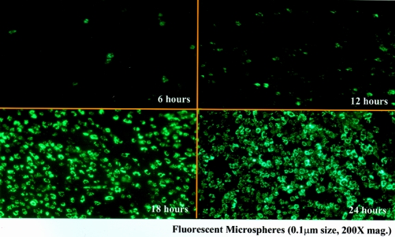
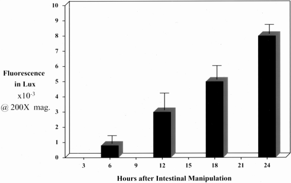
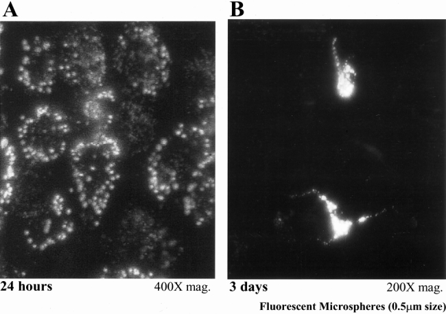
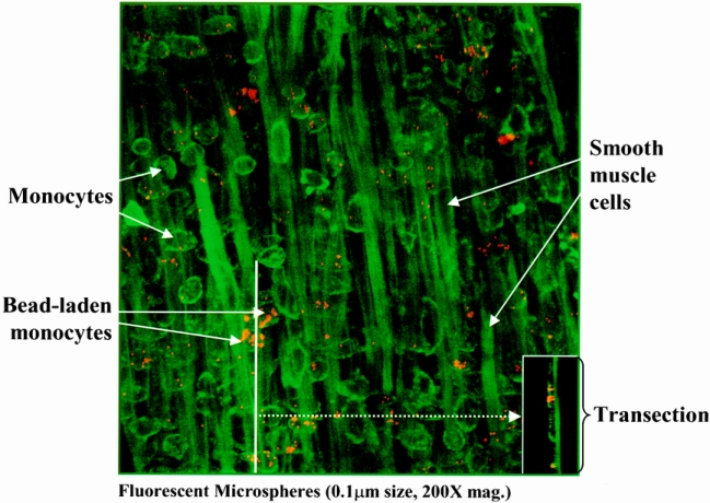
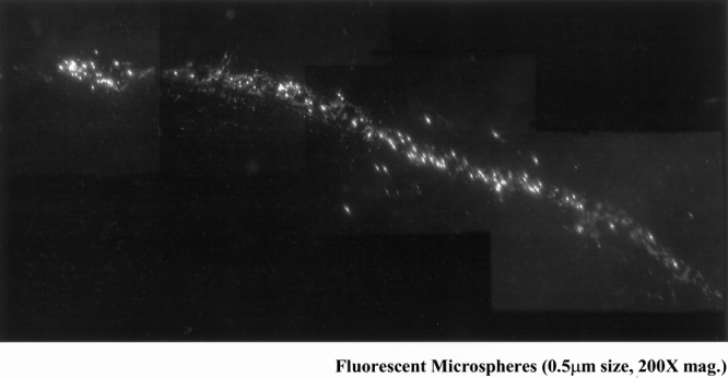
Methods: ACI rats were subjected to small bowel manipulation, after which fluorescent carboxylated or paramagnetic microspheres were administered into the gut lumen. Animals were killed between 0 and 24 hours; unoperated rats served as controls.

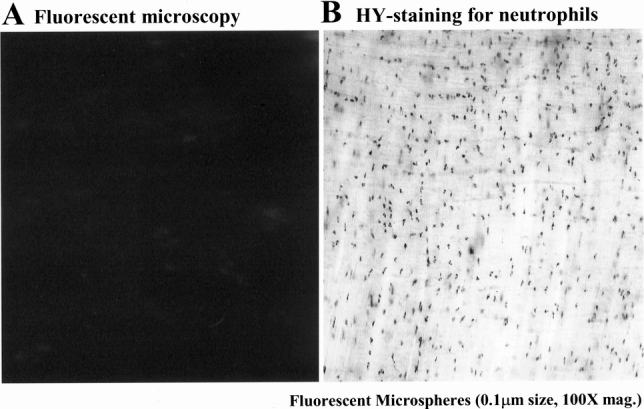
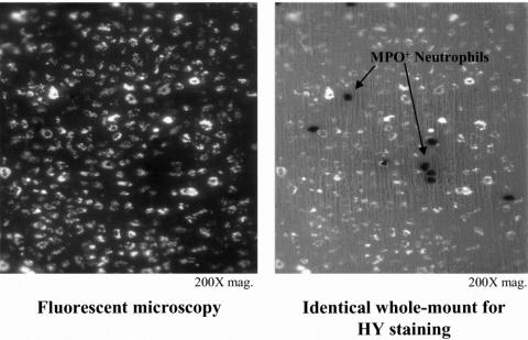
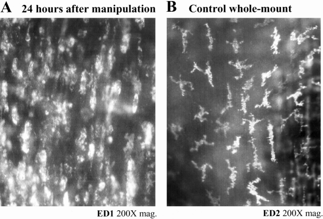
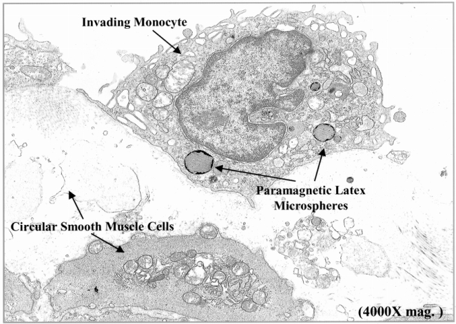
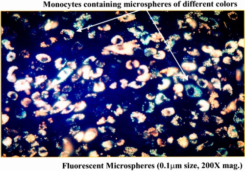
Results: Intestinal manipulation led to an early transient transference of microspheres from the intestinal lumen into mesenteric lymph that was not observed in unmanipulated controls. A time- dependent, significant increase in microsphere-laden phagocytes was observed within the intestinal muscularis. Immunohistochemistry and electron microscopy of the intestinal muscularis identified the phagocytes as extravasating ED1+ monocytes. Interruption of the lymphatics abolished the accumulation of microsphere-laden monocytes within the muscularis, although a significant monocytic recruitment could still be observed within the intestinal muscularis.

Conclusions: These data show that intestinal manipulation leads to a transient increase in mucosal permeability and that the extraintestinal endocytotic uptake of transferred particles by circulating monocytes precedes their infiltration into the gut wall. The transference of luminal bacterial products may follow a similar route and time course as the microspheres. The authors hypothesize that endogenous bacterial products act synergistically with the inflammatory response within the postsurgical intestinal muscularis, leading to an exacerbation of postoperative ileus.


Figures

None
Figure 1. Jejunal muscularis layers at different time points after intestinal manipulation of the rat small intestine with the intraluminal administration of fluorescent microspheres 0.1 μm in diameter. (Upper left) Starting at 6 hours after manipulation, cells containing fluorescent microspheres begin to be observed within muscularis externa whole mounts. This recruitment time-dependently increased until at 24 hours after intestinal manipulation a virtual carpet of microsphere-laden cells was seen within the muscularis (200× original magnification).
None
Figure 10. Subdiaphragmatic common lymph duct diversion prevented the appearance of fluorescent microspheres within phagocytes, which had extravasated into the manipulated jejunal muscularis. (Panel A) The muscularis whole mount from an animal in which the common subdiaphragmatic lymph duct was diverted by cutting after intestinal manipulation and microsphere injection showed the presence of no fluorescent beads in the muscularis (100× original magnification). However, these animals continued to have a strong leukocytic infiltrate, as shown by the presence of numerous myeloperoxidase-positive neutrophils within the manipulated muscularis (panel B, 200× original magnification).
None
Figure 2. The histogram quantifies the time-dependent increase in fluorescent microsphere accumulation within manipulated jejunal muscularis whole mounts measured with a lux meter at a magnification of 200× at 6, 12, 18, and 24 hours after intestinal manipulation (n = 4).
None
Figure 3. Double-labeling of this manipulated whole mount after 24 hours shows that the microsphere-laden cells in panel A photographed under fluorescent lighting are not identified as the strongly myeloperoxidase neutrophils shown in panel B, photographed with partial transilluminating light (200× original magnification).
None
Figure 4. Manipulation induced recruitment of monocytes (panel A) to the already dense network of normally resident macrophages (panel B) within the jejunal muscularis. Twenty-four hours after intestinal manipulation, a large amount of ED1+ round monocytes can be stained lying within the intestinal jejunal muscularis (A). A dense network of ED2+ macrophages could be discriminated in muscularis externa whole mounts from controls using a highly selective monoclonal antibody (B).
None
Figure 5. Morphologic transformation of newly recruited microsphere-laden monocytes to resident macrophages. Panel A shows a typical whole mount from an animal 24 hours after manipulation, illustrating the typical round aspect of these microsphere-containing phagocytes (400×). The morphologic appearance of the microsphere-laden monocytes changed over a 3-day period by transforming into more dendritic stellate resident macrophages (panel B, 400×).
None
Figure 6. Electron microscopic confirmation of the microsphere-laden phagocytes as monocytes lying within the manipulated jejunal muscularis. Jejunal muscularis preparations fixed in glutaraldehyde for electron microscopy show the accumulation of 0.46-μm paramagnetic latex beads in phagocytes, which had structural cellular characteristics of monocytes. Each monocyte generally contained several microspheres and could be observed to be engulfing necrotic smooth muscle cells (4,000× original magnification).
None
Figure 7. The postoperative distribution of microsphere-laden monocytes within the manipulated muscularis externa was investigated using confocal microscopy. The composite of the planar images shows numerous extravasated monocytes within jejunal muscularis 24 hours after intestinal manipulation. The transection composite (insert) shows the predominant localization of the fluorescently labeled monocytes near the serosa surface of the muscularis (panel, 200× magnification).
None
Figure 8. Coalescence within monocytes of two different-colored microspheres coming from two separate and independent loops of the manipulated small intestine. A two-loop small intestinal model was created to investigate the luminal transference pathway of the microspheres. The selective injection of yellow and red microspheres into individual intestinal loops and their combined appearance within extravasated monocytes within a single muscularis whole mount indicated that the microspheres do not transmurally pass through the gut wall to the muscularis, but that a more indirect extraintestinal route is taken.
None
Figure 9. The early appearance of fluorescent microspheres within the mesenteric lymph vessels could be observed within 4 hours after intestinal manipulation and bead administration. This figure shows a four-picture composite of microspheres draining through a mesenteric lymph vessel 4 hours after intestinal manipulation (200× original magnification).

Similar articles

Cited by

References

    1. Bates DW, Sands K, Miller E, et al. Predicting bacteremia in patients with sepsis syndrome. J Infect Dis 1997; 176: 1538–1551. - PubMed
    1. Livingston DH, Mosenthal AC, Deitch EA. Sepsis and multiple organ dysfunction syndrome: a clinical-mechanistic overview. New Horizons 1995; 3: 257–266. - PubMed
    1. Pape H-C, Dwenger A, Regel G, et al. Increased gut permeability after multiple trauma. Br J Surg 1994; 81: 850–852. - PubMed
    1. Saadia R. Trauma and bacterial translocation. Br J Surg 1995; 82: 1243–1244. - PubMed
    1. O’Boyle C, MacFie J, Mitchell CJ. The microbiology of bacterial translocation in humans. Gut 1998; 42: 29–35. - PMC - PubMed

Publication types