

Connective tissue growth factor is involved in pancreatic repair and tissue remodeling in human and rat acute necrotizing pancreatitis
- PMID: 11753043
- PMCID: PMC1422396
- DOI: 10.1097/00000658-200201000-00008
Connective tissue growth factor is involved in pancreatic repair and tissue remodeling in human and rat acute necrotizing pancreatitis
Abstract
Objective: To analyze the involvement of connective tissue growth factor (CTGF) in the transforming growth factor-beta (TGF-beta) pathway during acute necrotizing pancreatitis (ANP) in humans and rats.
Summary background data: Connective tissue growth factor is involved in several fibrotic diseases and has a critical role in fibrogenesis and tissue remodeling after injury.
Methods: Normal human pancreas tissue samples were obtained through an organ donor program from five individuals without a history of pancreatic disease. Human ANP tissues were obtained from eight persons undergoing surgery for this disease. In rats, ANP was induced by intraductal infusion of taurocholate. The expression of CTGF was studied by Northern blot analysis, in situ hybridization, and immunohistochemistry in both human and rat pancreatic tissue samples.
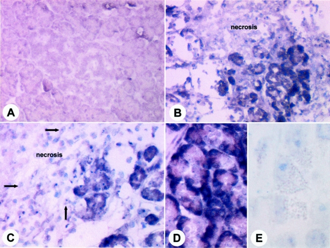
Results: Northern blot analysis revealed enhanced CTGF mRNA expression in human ANP tissue samples compared with normal controls. In addition, a concomitant increase in TGF-beta1 was present. By in situ hybridization, CTGF mRNA was localized in the remaining acinar and ductal cells and in fibroblasts. In regions of intense damage adjacent to areas of necrosis, CTGF mRNA signals were most intense. Inflammatory cells were devoid of any CTGF mRNA signals. By immunohistochemistry, CTGF protein was localized at high levels in the same cell types as CTGF mRNA. In ANP in rats, concomitantly enhanced mRNA levels of CTGF, TGF-beta1, and collagen type 1 were present, with a biphasic peak pattern on days 2 to 3 and day 7 after induction of ANP.
Conclusions: These data indicate that CTGF participates in tissue remodeling in ANP. The expression of CTGF predominantly in the remaining acinar and ductal cells indicates that extracellular matrix synthesis after necrosis is at least partly regulated by the remaining pancreatic parenchyma and only to a minor extent by inflammatory cells. Blockage of CTGF, a downstream mediator of TGF-beta in fibrogenesis, might be useful as a target to influence and reduce fibrogenesis in this disorder.
Figures


Similar articles
-
Transforming growth factor-beta pathway is activated in cholecystolithiasis.Langenbecks Arch Surg. 2005 Feb;390(1):21-8. doi: 10.1007/s00423-004-0517-4. Epub 2004 Nov 20. Langenbecks Arch Surg. 2005. PMID: 15702358
-
Connective tissue growth factor is a regulator for fibrosis in human chronic pancreatitis.Ann Surg. 1999 Jul;230(1):63-71. doi: 10.1097/00000658-199907000-00010. Ann Surg. 1999. PMID: 10400038 Free PMC article.
-
Connective tissue growth factor gene expression alters tumor progression in esophageal cancer.World J Surg. 2002 Apr;26(4):420-7. doi: 10.1007/s00268-001-0242-x. Epub 2002 Feb 4. World J Surg. 2002. PMID: 11910473
-
Connective tissue growth factor: a mediator of TGF-beta action on fibroblasts.Cytokine Growth Factor Rev. 1997 Sep;8(3):171-9. doi: 10.1016/s1359-6101(97)00010-5. Cytokine Growth Factor Rev. 1997. PMID: 9462483 Review.
-
Connective tissue growth factor: what's in a name?Mol Genet Metab. 2000 Sep-Oct;71(1-2):276-92. doi: 10.1006/mgme.2000.3059. Mol Genet Metab. 2000. PMID: 11001822 Review.
Cited by
-
Transforming growth factor-beta pathway is activated in cholecystolithiasis.Langenbecks Arch Surg. 2005 Feb;390(1):21-8. doi: 10.1007/s00423-004-0517-4. Epub 2004 Nov 20. Langenbecks Arch Surg. 2005. PMID: 15702358
-
Routine laboratory parameters in patients with necrotizing pancreatitis by the time of operative pancreatic debridement: Food for thought.World J Gastrointest Surg. 2022 Jan 27;14(1):64-77. doi: 10.4240/wjgs.v14.i1.64. World J Gastrointest Surg. 2022. PMID: 35126864 Free PMC article.
-
Connective tissue growth factor production by activated pancreatic stellate cells in mouse alcoholic chronic pancreatitis.Lab Invest. 2010 Aug;90(8):1179-88. doi: 10.1038/labinvest.2010.82. Epub 2010 Apr 5. Lab Invest. 2010. PMID: 20368699 Free PMC article.
-
Effect of early administration of exogenous basic fibroblast growth factor on acute edematous pancreatitis in rats.World J Gastroenterol. 2006 May 21;12(19):3060-4. doi: 10.3748/wjg.v12.i19.3060. World J Gastroenterol. 2006. PMID: 16718788 Free PMC article.
-
Activation of the intrinsic fibroinflammatory program in adult pancreatic acinar cells triggered by Hippo signaling disruption.PLoS Biol. 2019 Sep 12;17(9):e3000418. doi: 10.1371/journal.pbio.3000418. eCollection 2019 Sep. PLoS Biol. 2019. PMID: 31513574 Free PMC article.
References
-
- Menke A, Yamaguchi H, Giehl K, et al. Hepatocyte growth factor and fibroblast growth factor are over-expressed after cerulein-induced acute pancreatitis. Pancreas 1999; 18: 28–33. - PubMed
-
- Ebert M, Yokoyama M, Ishiwata T, et al. Alteration of fibroblast growth factor and receptor expression after acute pancreatitis in humans. Pancreas 1999; 18: 240–246. - PubMed
-
- Ludwig CU, Menke A, Adler G, et al. Fibroblasts stimulate acinar cell proliferation through IGF-I during regeneration from acute pancreatitis. Am J Physiol 1999; 276: G193–198. - PubMed
Publication types
MeSH terms
Substances
LinkOut - more resources
Full Text Sources
Miscellaneous
