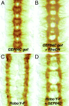
A novel Dbl family RhoGEF promotes Rho-dependent axon attraction to the central nervous system midline in Drosophila and overcomes Robo repulsion
- PMID: 11756465
- PMCID: PMC2199320
- DOI: 10.1083/jcb.200110077
A novel Dbl family RhoGEF promotes Rho-dependent axon attraction to the central nervous system midline in Drosophila and overcomes Robo repulsion
Abstract
The key role of the Rho family GTPases Rac, Rho, and CDC42 in regulating the actin cytoskeleton is well established (Hall, A. 1998. Science. 279:509-514). Increasing evidence suggests that the Rho GTPases and their upstream positive regulators, guanine nucleotide exchange factors (GEFs), also play important roles in the control of growth cone guidance in the developing nervous system (Luo, L. 2000. Nat. Rev. Neurosci. 1:173-180; Dickson, B.J. 2001. Curr. Opin. Neurobiol. 11:103-110). Here, we present the identification and molecular characterization of a novel Dbl family Rho GEF, GEF64C, that promotes axon attraction to the central nervous system midline in the embryonic Drosophila nervous system. In sensitized genetic backgrounds, loss of GEF64C function causes a phenotype where too few axons cross the midline. In contrast, ectopic expression of GEF64C throughout the nervous system results in a phenotype in which far too many axons cross the midline, a phenotype reminiscent of loss of function mutations in the Roundabout (Robo) repulsive guidance receptor. Genetic analysis indicates that GEF64C expression can in fact overcome Robo repulsion. Surprisingly, evidence from genetic, biochemical, and cell culture experiments suggests that the promotion of axon attraction by GEF64C is dependent on the activation of Rho, but not Rac or Cdc42.
Figures


Similar articles
-
Cross GTPase-activating protein (CrossGAP)/Vilse links the Roundabout receptor to Rac to regulate midline repulsion.Proc Natl Acad Sci U S A. 2005 Mar 22;102(12):4613-8. doi: 10.1073/pnas.0409325102. Epub 2005 Mar 8. Proc Natl Acad Sci U S A. 2005. PMID: 15755809 Free PMC article.
-
Dosage-sensitive and complementary functions of roundabout and commissureless control axon crossing of the CNS midline.Neuron. 1998 Jan;20(1):25-33. doi: 10.1016/s0896-6273(00)80431-6. Neuron. 1998. PMID: 9459439
-
Vilse, a conserved Rac/Cdc42 GAP mediating Robo repulsion in tracheal cells and axons.Genes Dev. 2004 Sep 1;18(17):2161-71. doi: 10.1101/gad.310204. Genes Dev. 2004. PMID: 15342493 Free PMC article.
-
Axon guidance: mice and men need Rig and Robo.Curr Biol. 2004 Aug 10;14(15):R632-4. doi: 10.1016/j.cub.2004.07.050. Curr Biol. 2004. PMID: 15296783 Review.
-
[Current progress in functions of axon guidance molecule Robo and underlying molecular mechanism].Sheng Li Xue Bao. 2014 Jun 25;66(3):373-85. Sheng Li Xue Bao. 2014. PMID: 24964856 Review. Chinese.
Cited by
-
Rho-guanine nucleotide exchange factors during development: Force is nothing without control.Small GTPases. 2010 Jul;1(1):28-43. doi: 10.4161/sgtp.1.1.12672. Small GTPases. 2010. PMID: 21686118 Free PMC article.
-
Drosophila as a genetic and cellular model for studies on axonal growth.Neural Dev. 2007 May 2;2:9. doi: 10.1186/1749-8104-2-9. Neural Dev. 2007. PMID: 17475018 Free PMC article. Review.
-
A vascular gene trap screen defines RasGRP3 as an angiogenesis-regulated gene required for the endothelial response to phorbol esters.Mol Cell Biol. 2004 Dec;24(24):10515-28. doi: 10.1128/MCB.24.24.10515-10528.2004. Mol Cell Biol. 2004. PMID: 15572660 Free PMC article.
-
Redundant mechanisms for regulation of midline crossing in Drosophila.PLoS One. 2008;3(11):e3798. doi: 10.1371/journal.pone.0003798. Epub 2008 Nov 24. PLoS One. 2008. PMID: 19030109 Free PMC article.
-
Small GTPase Cdc42 is required for multiple aspects of dendritic morphogenesis.J Neurosci. 2003 Apr 15;23(8):3118-23. doi: 10.1523/JNEUROSCI.23-08-03118.2003. J Neurosci. 2003. PMID: 12716918 Free PMC article.
References
-
- Awasaki, T., M. Saito, M. Sone, E. Suzuki, R. Sakai, K. Ito, and C. Hama. 2000. The Drosophila trio plays an essential role in patterning of axons by regulating their directional extension. Neuron. 26:119–131. - PubMed
-
- Bashaw, G.J., and C.S. Goodman. 1999. Chimeric axon guidance receptors: the cytoplasmic domains of slit and netrin receptors specify attraction versus repulsion. Cell. 97:917–926. - PubMed
-
- Bashaw, G.J., T. Kidd, D. Murray, T. Pawson, and C.S. Goodman. 2000. Repulsive axon guidance: Abelson and Enabled play opposing roles downstream of the roundabout receptor. Cell. 101:703–715. - PubMed
-
- Bateman, J., H. Shu, and D. Van Vactor. 2000. The guanine nucleotide exchange factor trio mediates axonal development in the Drosophila embryo. Neuron. 26:93–106. - PubMed
-
- Cerione, R.A., and Y. Zheng. 1996. The Dbl family of oncogenes. Curr. Opin. Cell Biol. 8:216–222. - PubMed
Publication types
MeSH terms
Substances
Grants and funding
LinkOut - more resources
Full Text Sources
Molecular Biology Databases
Miscellaneous

