. 2001 Dec 24;155(7):1137-45.
doi: 10.1083/jcb.200109063. Epub 2001 Dec 24.

Implication of a novel multiprotein Dam1p complex in outer kinetochore function

Affiliations

Implication of a novel multiprotein Dam1p complex in outer kinetochore function

I M Cheeseman et al. J Cell Biol. .

Abstract

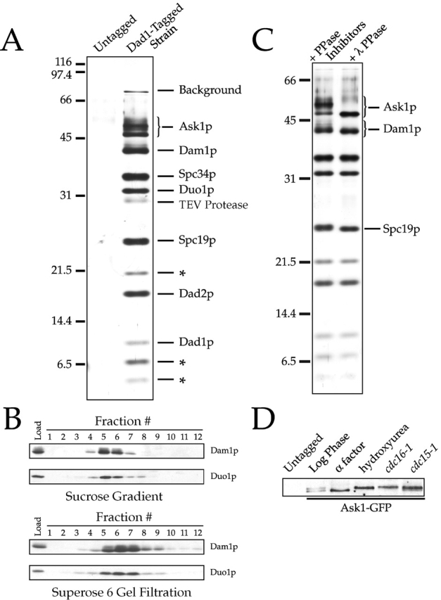
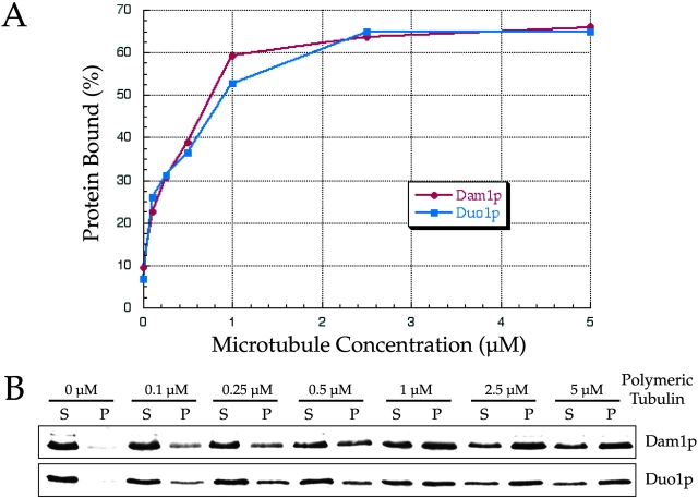
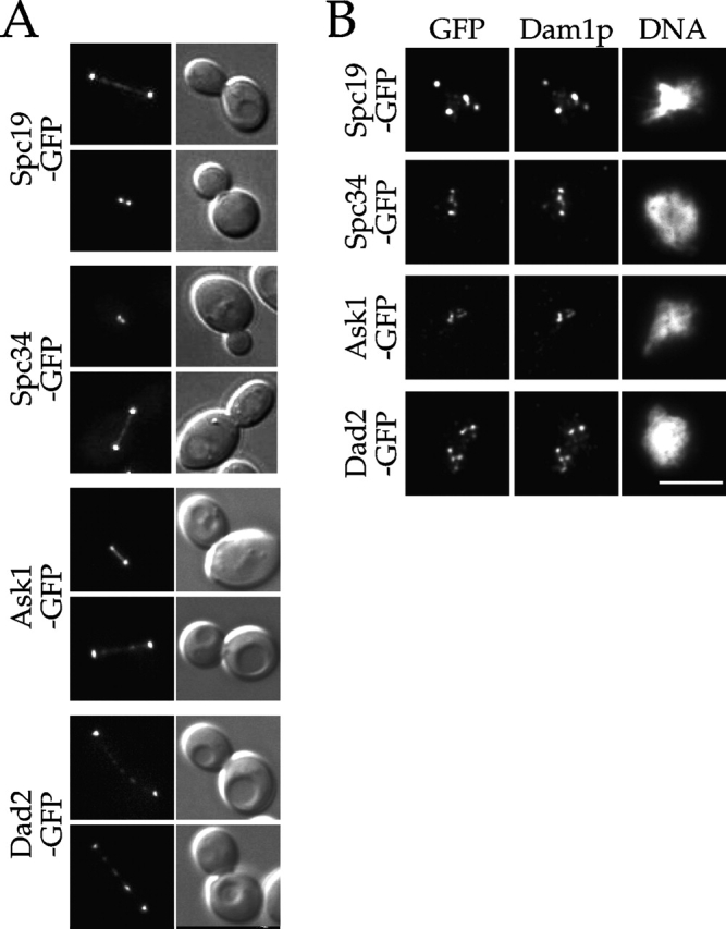
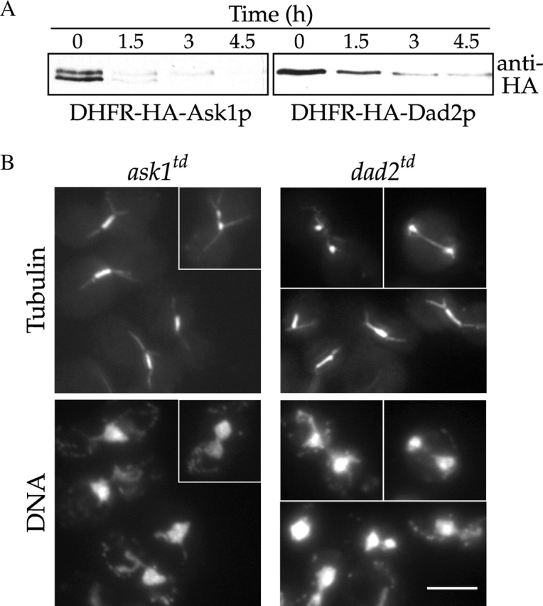
Dam1p, Duo1p, and Dad1p can associate with each other physically and are required for both spindle integrity and kinetochore function in budding yeast. Here, we present our purification from yeast extracts of an approximately 245 kD complex containing Dam1p, Duo1p, and Dad1p and Spc19p, Spc34p, and the previously uncharacterized proteins Dad2p and Ask1p. This Dam1p complex appears to be regulated through the phosphorylation of multiple subunits with at least one phosphorylation event changing during the cell cycle. We also find that purified Dam1p complex binds directly to microtubules in vitro with an affinity of approximately 0.5 microM. To demonstrate that subunits of the Dam1p complex are functionally important for mitosis in vivo, we localized Spc19-green fluorescent protein (GFP), Spc34-GFP, Dad2-GFP, and Ask1-GFP to the mitotic spindle and to kinetochores and generated temperature-sensitive mutants of DAD2 and ASK1. These and other analyses implicate the four newly identified subunits and the Dam1p complex as a whole in outer kinetochore function where they are well positioned to facilitate the association of chromosomes with spindle microtubules.


Figures

Figure 1.
Figure 1.
Purification of a 245 kD Dam1p complex. (A) Purification of the Dam1p complex using a tagged Dad1p reveals 10 polypeptides. Purified Dam1p complex (as described in Materials and methods) and protein from an identical purification using an untagged control strain were separated on a 13.5% SDS-PAGE gel and silver stained. Polypeptides identified by mass spectrometric analysis of the complex are indicated. Those not yet identified are denoted by an asterisk. Bands labeled “background” are the highly homologous heat shock proteins Ssb1 and Ssb2 (66 kD) and Ssa1, Ssa2, Ssa3, and Ssa4 (70 kD). (B) Determination of S value and Stoke's radius of the Dam1p complex in yeast extracts. Sucrose gradient and Superose 6 gel filtration fractions (as described in Materials and methods) were probed with antibodies against Duo1p and Dam1p. The S value for Duo1p/Dam1p-containing complex was estimated as 6.5 from a linear fit of the S values versus peak fraction number of standards, and the Stoke's radius was estimated as 90.1 Å from a Porath correlation, relating to the elution volumes of the standard proteins to their known Stoke's radii (Siegel and Monty, 1966). (C) Multiple subunits of the Dam1p complex are phosphorylated in vivo. Dam1p complex was purified in the presence of phosphatase inhibitors and then split into two aliquots, one of which was treated with lambda protein phosphatase. (D) Ask1p phosphorylation changes over the cell cycle. Ask1-GFP–tagged strains were grown to log phase and arrested in either α-factor (G1), 0.2 M hydroxyurea (S phase), or by using temperature-sensitive cdc16–1 (metaphase) and cdc15–1 (telophase) mutants. Protein samples were run on an 8% SDS-PAGE gel and immunoblotted with antibodies against GFP.
Figure 2.
Figure 2.
Purified Dam1p complex binds to microtubules directly with0.5 μM affinity. Purified Dam1p complex (∼5–10 nM) was incubated with varying concentrations of microtubules, which were then pelleted by centrifugation. The amount of complex bound to microtubules was determined by quantifying Duo1p and Dam1p in the pellet and supernatant fractions. (A) Percentage of protein bound to microtubules plotted with respect to the concentration of microtubules in the reaction. (B) Western blots showing the amount of Dam1p or Duo1p that is unbound (S, supernatant) or bound (P, pellet) at each concentration of microtubules.
Figure 3.
Figure 3.
Spc19p, Spc34p, Dad2p, and Ask1p localize to spindles and kinetochores. (A) GFP fluorescence and corresponding DIC images showing the localization of the indicated fusion protein to the mitotic spindle. (B) Cells expressing the indicated GFP fusion proteins were prepared for chromosome spreads as described (Loidl et al., 1998). They were then processed for immunofluorescence and stained with anti-GFP and anti-Dam1p antibodies. Bar, 5 μm.
Figure 4.
Figure 4.
dad2 and ask1 mutants show spindle defects. (A) Immunoblots of ask1 td and dad2 td strains showing degradation of the fusion protein. Degron-tagged alleles of ask1 and dad2 were grown at 25°C and shifted to 37°C at t = 0 h Protein samples were immunoblotted with anti-HA antibodies to detect the DHFRts–HA fusion protein. (B) ask1 td and dad2 td mutant phenotypes. Degron-tagged alleles of ask1 and dad2 were grown at 25°C and shifted to 37°C for 3 h. They were then processed for tubulin immunofluorescence and DNA staining (DAPI). At this time point, 90% of large budded ask1 td and 82% of large budded dad2 td cells showed short spindle structures and a single mass of DNA, whereas 10% of ask1 td and 15% of dad2 td cells showed broken down spindles (n = 100 cells/sample). Bar, 5 μm.
Figure 5.
Figure 5.
Analysis of stu2 dam1 and dam1 bik1 double mutants reveals shared roles in spindle structure. dam1-11, stu2-10, and bik1-1 single mutants and dam1-11 stu2-10, dam1-1 stu2-10, dam1-11 bik1-1, and dam1-1 bik1-1 double mutants were grown at 25°C and shifted to 37°C for 3 h. They were then processed for tubulin immunofluorescence and DNA staining (DAPI). Bar, 5 μm.
Figure 6.
Figure 6.
Protein interactions establishing the connectivity between spindle microtubules and centromeric DNA. Physical interactions are indicated by lines between proteins. Shapes indicate distinct complexes within the kinetochore. Proteins that interact directly with centromeric DNA are shown in blue. Proteins whose loss of function results in genetic interactions with dam1-1 are shown in red. Protein interaction data are from Chen et al. (1998), Ghosh et al. (2001), Ito et al. (2001), Ito et al. (2000), Newman et al. (2000), Ortiz et al. (1999), Uetz et al. (2000), Yoon and Carbon (1999), and Kang et al. (2001). Physical interactions between Mcm16-Ctf19, Mcm22-Ctf19, Mcm16-Ctf3, Mcm22-Ctf3, and Spc34-Mcm22 represent coimmunoprecipitation and two-hybrid data from V. Measday and P. Hieter (personal communication).

Similar articles

Cited by

References

    1. Ayscough, K.R., and D.G. Drubin. 1998. Immunofluorescence microscopy of yeast cells. Cell Biology: A Laboratory Handbook. Vol. 2. J. Celis, editor. Academic Press, Inc., New York. 477–485.
    1. Cheeseman, I.M., M. Enquist-Newman, T. Müller-Reichert, D.G. Drubin, and G. Barnes. 2001. Mitotic spindle integrity and kinetochore function linked by the Duo1p/Dam1p complex. J. Cell Biol. 152:197–212. - PMC - PubMed
    1. Chen, X.P., H. Yin, and T.C. Huffaker. 1998. The yeast spindle pole body component Spc72p interacts with Stu2p and is required for proper microtubule assembly. J. Cell Biol. 141:1169–1179. - PMC - PubMed
    1. Dohmen, R.J., P. Wu, and A. Varshavsky. 1994. Heat-inducible degron: a method for constructing temperature-sensitive mutants. Science. 263:1273–1276. - PubMed
    1. Enquist-Newman, M., I.M. Cheeseman, D. Van Goor, D.G. Drubin, P. Meluh, and G. Barnes. 2001. Dad1p, third component of the Duo1p/Dam1p complex involved in kinetochore function and mitotic spindle integrity. Mol. Biol. Cell. 12:2601–2613. - PMC - PubMed

Publication types

MeSH terms