. 2001 Dec 24;155(7):1185-98.
doi: 10.1083/jcb.200105102. Epub 2001 Dec 24.

Abelson kinase regulates epithelial morphogenesis in Drosophila

Affiliations

Abelson kinase regulates epithelial morphogenesis in Drosophila

E E Grevengoed et al. J Cell Biol. .

Abstract

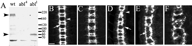
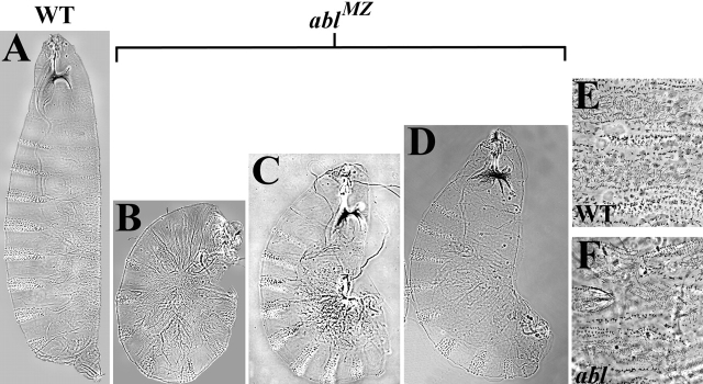
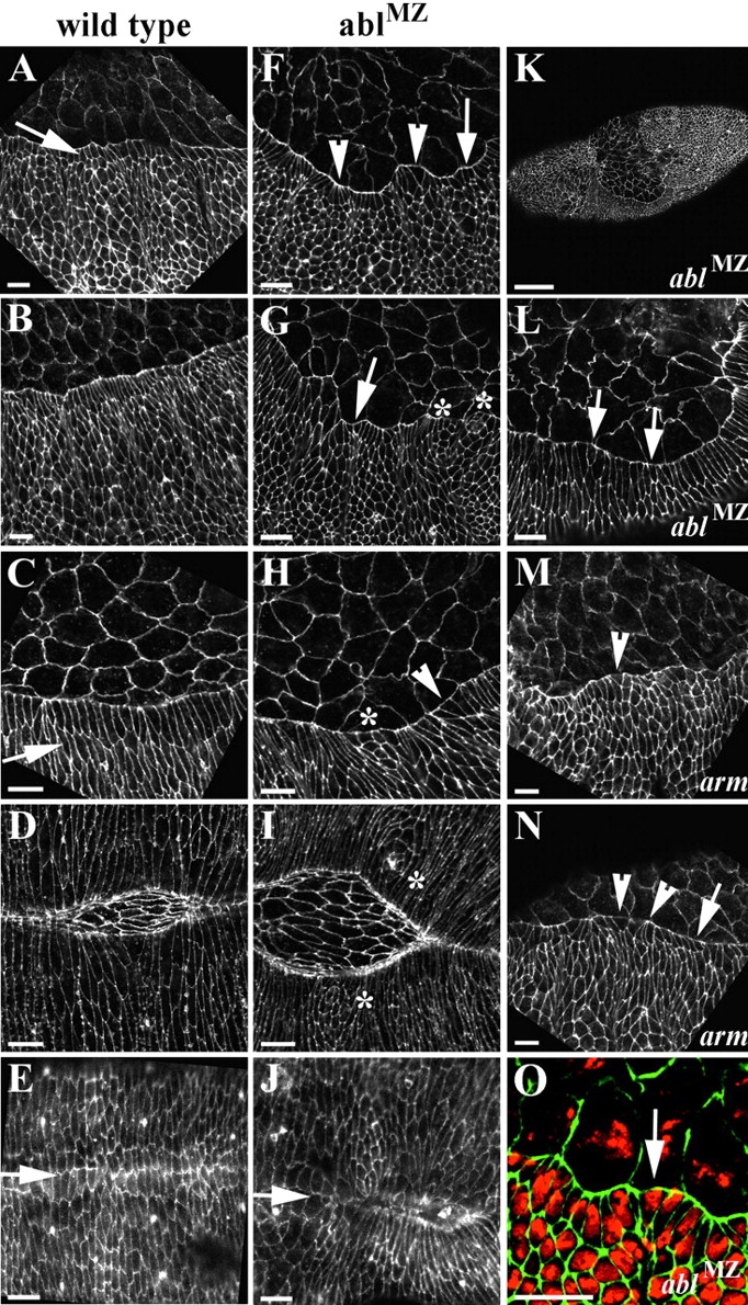
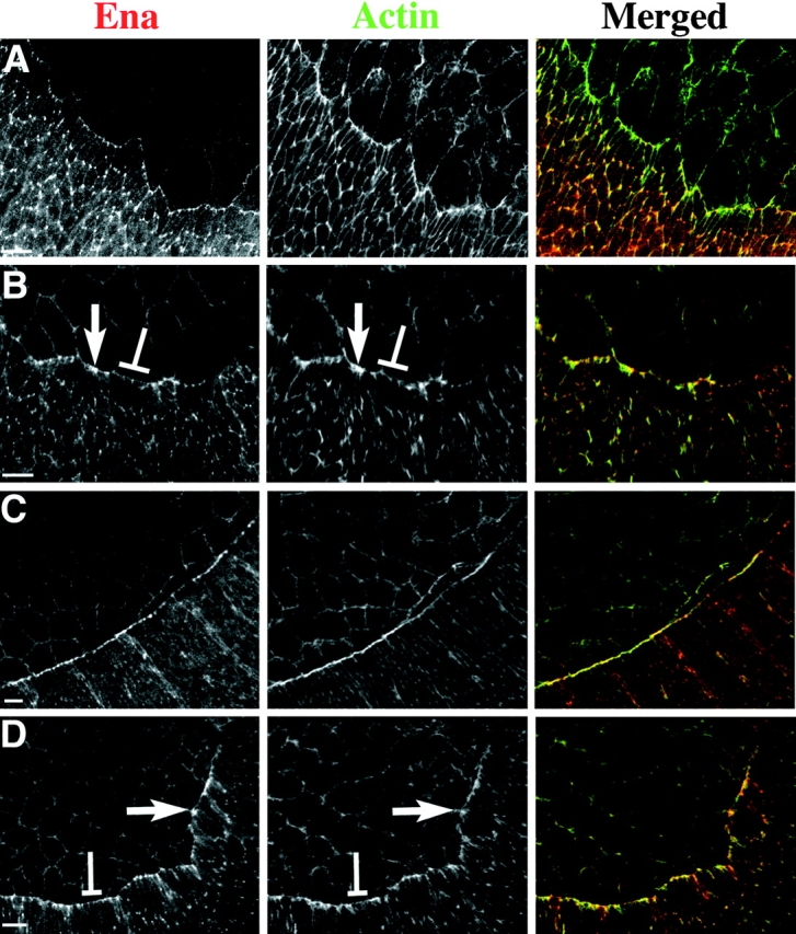
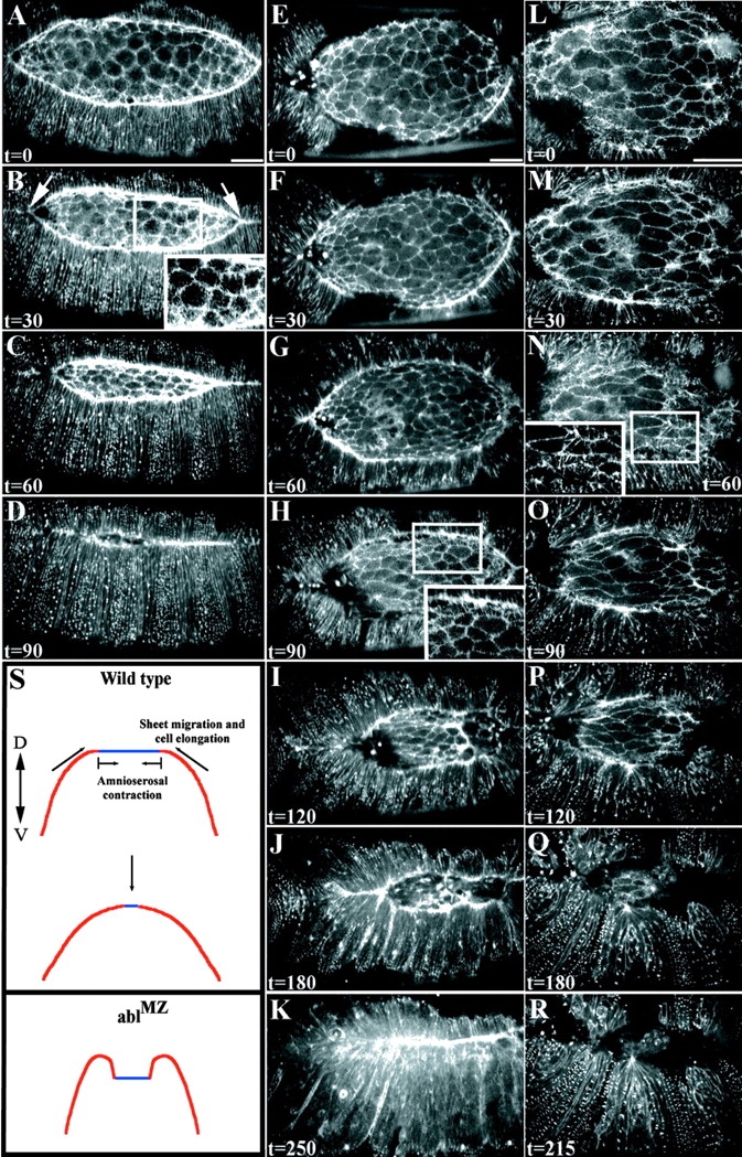
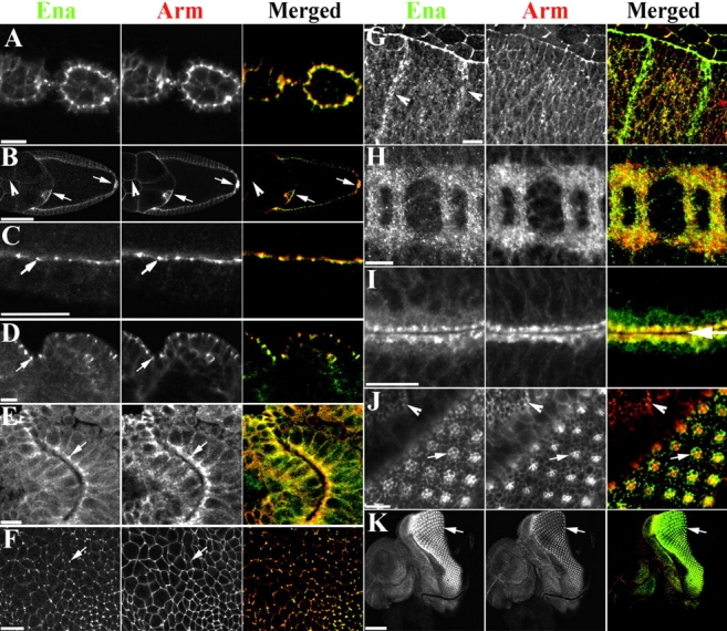
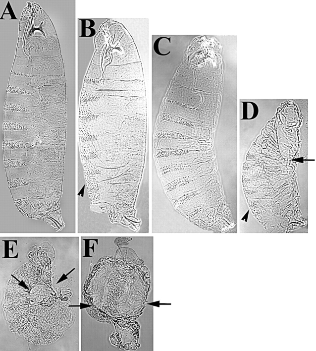
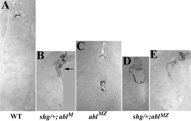
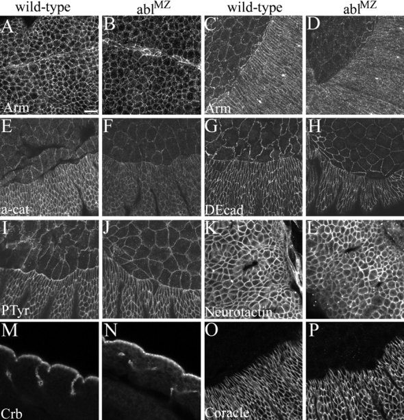
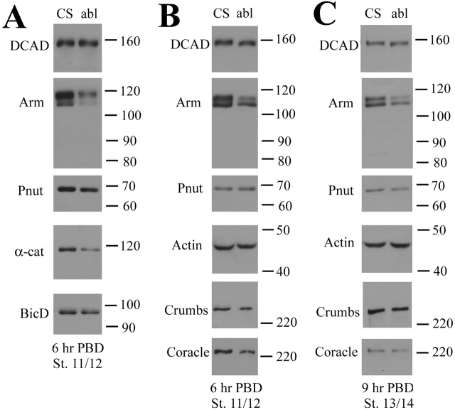
Activation of the nonreceptor tyrosine kinase Abelson (Abl) contributes to the development of leukemia, but the complex roles of Abl in normal development are not fully understood. Drosophila Abl links neural axon guidance receptors to the cytoskeleton. Here we report a novel role for Drosophila Abl in epithelial cells, where it is critical for morphogenesis. Embryos completely lacking both maternal and zygotic Abl die with defects in several morphogenetic processes requiring cell shape changes and cell migration. We describe the cellular defects that underlie these problems, focusing on dorsal closure as an example. Further, we show that the Abl target Enabled (Ena), a modulator of actin dynamics, is involved with Abl in morphogenesis. We find that Ena localizes to adherens junctions of most epithelial cells, and that it genetically interacts with the adherens junction protein Armadillo (Arm) during morphogenesis. The defects of abl mutants are strongly enhanced by heterozygosity for shotgun, which encodes DE-cadherin. Finally, loss of Abl reduces Arm and alpha-catenin accumulation in adherens junctions, while having little or no effect on other components of the cytoskeleton or cell polarity machinery. We discuss possible models for Abl function during epithelial morphogenesis in light of these data.


Figures

Figure 1.
Figure 1.
Complete loss of Abl disrupts CNS development. (A) Cell extracts from 3-h-old wild-type embryos or embryonic progeny of females with abl mutant germlines, immunoblotted with anti-Abl antibody. Wild-type Abl is ∼180 kD (top arrow). abl 4 does not produce a protein recognized by this antibody. abl 1 produces a truncated protein product of ∼80 kD (bottom arrow). (B–F) Embryos (anterior up) labeled with mAb BP102, which labels all axons. (B) Wild-type CNS, with a scaffold of longitudinal (arrowhead) and commissural (arrow) axons. (C and D) Maternally abl mutant but zygotically-rescued embryos have relatively wild-type CNS development, with occasional collapsed longitudinal axons (D, arrowhead) and gaps in axon bundles (D, arrow). (E and F) abl MZ mutants exhibit severe disruptions in CNS development. Most exhibit loss of commissural axons (arrow, E) and some have defects in both longitudinal and commissural axons (F). Bar, 25 μm.
Figure 2.
Figure 2.
ablMZ mutants have defects in epithelial morphogenesis. Cuticle preparations, anterior up. In A–D, dorsal is to the right. (A) Wild-type. (B–D) The range of phenotypes in abl4 maternal/zygotic mutants, a similar range is observed in abl 1 . (B) Approximately 7% of abl MZ mutants have head involution defects and completely fail to germband retract. (C) ∼14% of abl MZ mutants partially fail to germband retract and have variable dorsal closure defects. Note the dorsal hole (arrow). (D) Approximately 67% of abl MZ mutants have dorsal closure defects. (E) Wild-type dorsal hair pattern. (F) Misaligned dorsal hairs in abl MZ mutant.
Figure 3.
Figure 3.
ablMZ mutants fail to undergo coordinated changes in cell shape during dorsal closure. Embryos labeled with antiphosphotyrosine. Anterior is to the left. (A–E) Wild-type at progressively later stages of dorsal closure. (A–C) Lateral views. (D and E) Dorsal views. (A) Leading edge cells have begun to uniformly elongate (arrow). (B and C) Successive lateral cell rows uniformly elongate (arrow). (D) Lateral epithelial sheets zip together. (E) Closure is complete, with lateral epithelial cells evenly matched at the midline (arrow). (F–J) abl MZ mutants at progressively later stages of dorsal closure. (F–H) Lateral and (I–J) dorsal views. (F) Leading edge cells do not elongate uniformly (arrow). Some cells have broadened or constricted leading edges (arrowheads). (G and H) Lateral cells have begun to elongate, but do so nonuniformly (arrow). Some cells have broadened or narrowed leading edges (arrowheads). Other groups of cells completely fail to elongate (asterisks). (I) abl MZ mutants that proceeded through dorsal closure. Small groups of cells have still completely failed to change shape (asterisks). (J) abl MZ mutant that completed closure. Epithelial sheets often fail to align properly at the midline (arrow). (K and L) Some abl MZ mutants initiate dorsal closure even though they have not completed germband retraction. Cell shape defects are also seen in these embryos (arrows). (M and N) arm XP33 mutants have cell shape defects similar to abl MZ mutants. Cells fail to elongate uniformly (arrow) and have broadened or narrowed leading edges (arrowheads). (O) Cell shape defects in abl MZ mutants are not caused by multinucleate cells. abl MZ, double-labeled with antiphosphotyrosine and with propidium iodide, labeling nuclei. Mononucleate cells have defects in shape (arrow). Bars: (A–J and L–O) 10 μm; (K) 50 μm.
Figure 4.
Figure 4.
Complete loss of Abl alters Ena and actin localization during dorsal closure. Lateral view. Embryos double-labeled with anti-Ena (red) and phalloidin (green), labeling F-actin. (A) Stage 13 wild-type embryo with leading edge cells initiating elongation. Ena (left, red) is enriched at vertices of cell–cell contact. Actin (middle, green) outlines all cell membranes and is beginning to accumulate at the leading edge. Actin and Ena colocalize at cell junctions. (B) Stage 13 abl MZ mutant. Ena (left, red) and Actin (middle, green) enrichment is not uniform at the leading edge. Both are enriched in some cells (arrows) and depleted in others (brackets). (C) Stage 14 wild-type embryo. More lateral cells have undergone uniform elongation. Ena is uniformly enriched at adherens junctions of leading edge cells (left, red). Actin forms a tight cable along the leading edge (middle, green). Ena and actin colocalize at adherens junctions as actin expands along the entire leading edge. (D) Stage 14 abl MZ mutant. Nonuniform localization of Ena and Actin persists. Cells with excess Ena (left, arrow) often accumulate excess Actin (middle, arrow), whereas cells with diminished Ena levels (left, bracket) have diminished levels of Actin (middle, bracket) Bars, 10 μm.
Figure 5.
Figure 5.
Dorsal closure is substantially slowed in ablMZ mutants. Dorsal view of embryos expressing moesin-GFP, anterior to the right. Time is in minutes. Insets in (B, H, and N) display actin-rich filopodia extending from amnioserosa or leading edge cells. (A–D) Wild-type embryo at 30 min intervals during dorsal closure. (A) The leading edge of the dorsal closure front is uniformly enriched in actin. Lateral epithelial sheets elongate uniformly. (B) 30 min. Amnioserosa cells are undergoing apical constriction and the embryo is zipping together at the anterior and posterior ends (arrows). (C) 60 min. Amnioserosa cells have constricted apically and remain in the same plane of focus as the lateral epithelial sheets. (D) 90 min. Dorsal closure is complete. (E–K) abl MZ mutant. The amnioserosa cells in E are comparable in surface area to the wild-type in A. (F–H) As closure progresses, amnioserosa cells constrict and lateral epithelial cells elongate, but dorsal closure is delayed relative to wild-type (compare D and H). If one matches embryos based on the length of the leading edge (compare A and G), closure is still delayed. This embryo took >4 h to complete closure (K). (L–R) A different abl MZ mutant at higher magnification, illustrating the folding-under of the leading edge and failure to complete closure. (S) Cross-section diagram depicting one interpretation of the defects of abl MZ mutants. In wild-type embryos, the rate at which lateral cells elongate, the sheets migrate, and amnioserosa cells constrict are tightly coordinated. In abl MZ mutants, the leading edge folds under the more lateral epidermis, perhaps because leading edge cells migrate too slowly or amnioserosa cell constriction is slowed (these events are likely coupled), forcing the sheet to fold under. Time-lapse videos supplementing this figure are available at http://www.jcb.org/cgi/content/full/jcb.200105102/DC1. Bars, 25 μm.
Figure 6.
Figure 6.
Ena and Arm colocalize at adherens junctions throughout development. All images (except I) are anterior to the right. Ena is green and Arm is red in merged images. (A) Stage 3 egg chamber. Ena and Arm are enriched in apical adherens junctions of follicle cells. (B and C) Stage 10 egg chamber. Levels of Ena and Arm drop, but both remain at adherens junctions. Anterior (border cells) and posterior polar follicle cells are enriched in both Ena and Arm (B, arrows). Ena and Arm also colocalize to nurse cell membranes (B, arrowhead). (D–G) During embryogenesis Ena and Arm colocalize to adherens junctions of epithelial tissues. (D) Cross-section, stage 9 embryo. Ectodermal adherens junctions (arrow). (E) Adherens junctions of polarized cells of the invaginating hindgut (arrow). (F) Apical view, stage 9 embryo. Ena is enriched at vertices of cell–cell contact (arrow), whereas Arm is more uniform. (G) During dorsal closure Ena is enriched at adherens junctions of leading edge cells, but it is also found in the cytoplasm of cells at the segment boundary (arrowheads). (H) Ena and Arm both localize to axons. (I–K) Imaginal discs. (I) Apical surfaces of two epithelial sheets opposed to one another in the wing imaginal disc (arrow). Ena and Arm colocalize to apical adherens junctions, and are also found at the apical surface. (J and K) In eye imaginal discs cell differentiation occurs after the morphogenetic furrow passes. In undifferentiated cells, Ena and Arm colocalize to cell boundaries (J, arrowhead). As groups of cells begin differentiating as photoreceptors (J, arrow), Ena localizes uniformly to all cells of the precluster. Arm, in contrast, accumulates at high levels in a subset of these cells. Later, Ena and Arm colocalize in photoreceptor rhabdomeres (K, arrow). Bars: (A–J) 10 μm; (K) 50 μm.
Figure 7.
Figure 7.
Mutations in ena enhance arm's dorsal closure defects. Cuticle preparations, anterior up. (A) Wild-type. (B) arm H8.6 mutants have segment polarity defects due to defects in Wingless signaling (arrowhead), but head involution and dorsal closure are normal. (C) ena 210/ena GC1. (D) arm H8.6/Y; ena GC1/ena GC1. Note strong defects in dorsal closure and head involution (arrow), with no change in the segment polarity phenotype (arrowhead). (E) arm YD35 mutants have a dorsal hole (arrows), as well strong segment polarity defects. (F) The dorsal side of arm YD35; ena 210/ena 210 embryos is completely open (arrows).
Figure 8.
Figure 8.
DE-cadherin (shg) genetically interacts with Abl. Cuticle preparations, anterior up. (A) Wild-type. (B) abl 4/abl 4 × shg 2/CyO. abl 4 maternal mutants are zygotically rescued, with all hatching as larvae and most appearing wild-type. shg heterozygosity prevents zygotic rescue of abl maternal mutants and leads to morphogenesis defects. Note defects in dorsal closure and head involution (arrow). (C) abl MZ mutants die during embryogenesis with defects in epithelial morphogenesis. (D and E) shg heterozygosity enhances the abl MZ phenotype. (D) 70% of lethal progeny of abl 4/abl 4 × shg 2/+; abl 4/+ have cuticles that are reduced in size with a large dorsal-anterior hole. (E) 30% of the lethal progeny of abl 4/abl 4 × shg R69/+; abl 4/+ have a prominent dorsal-anterior hole.
Figure 9.
Figure 9.
Levels of junctional Arm and α-catenin are reduced in ablMZ mutants. Embryos labeled with anti-Arm (A–D), anti–α-catenin (E and F), anti–DE-cadherin (G and H), antiphosphotyrosine (I and J), anti-Neurotactin (K and L), anti-Crumbs (M and N), and anti-Coracle (O-P). (A and B) Stage 8. (C and D) Stage 14. (A and C) Wild-type. Arm localizes to adherens junctions. (B and D) abl MZ mutants. Arm accumulates at reduced levels in adherens junctions. (E and F) Stage 13. (E) Wild-type. α-catenin localizes to adherens junctions. (F) abl MZ mutant. α-catenin accumulates at reduced levels in adherens junctions. (G–J) Stage 13. DE-cadherin localizes to adherens junctions, without striking differences in localization or levels between wild-type (G) and abl MZ mutants (H). Phosphotyrosine localizes to adherens junctions, without noticeable differences between wild-type (I) and abl MZ mutants (J). (K and L) Stage 11. Neurotactin localizes to the baso-lateral membrane without noticeable differences between wild-type (K) and abl MZ mutants (L). (M and N) Cross-sectional views, Stage 11. Crumbs localizes to the apical membrane of epithelial cells without striking differences between wild-type (M) and abl MZ mutants (N). (O and P) Stage 13. Coracle localizes to septate junctions with no striking difference in levels between wild-type (O) and abl MZ mutants (P). Bar, 10 μm.
Figure 10.
Figure 10.
Total levels of Arm and α-catenin are reduced in abl mutants. abl germline mutant females were mated to abl heterozygous males and progeny were picked at the cellular blastoderm stage and aged for the indicated time postblastoderm (PBD). Wild-type embryos (Canton S [CS]) served as a control. Cell extracts were fractionated by SDS-PAGE and immunoblotted with the indicated antibodies. Molecular weight markers are to the right. Bicaudal D (BicD) or Peanut (Pnut) are loading controls. Each vertical set of samples represents sequential reprobing of the same blot. (A and B) Stage 11/12 embryos (6 h postblastoderm). (C) Stage 13/14 embryos (9 h postblastoderm).

Similar articles

Cited by

References

    1. Bashaw, G.J., T. Kidd, D. Murray, T. Pawson, and C.S. Goodman. 2000. Repulsive axon guidance: Abelson and Enabled play opposing roles downstream of the roundabout receptor. Cell. 101:703–715. - PubMed
    1. Baum, B., and N. Perrimon. 2001. Spatial control of the actin cytoskeleton in Drosophila epithelial cells. Nat. Cell Biol. 3:883–890. - PubMed
    1. Bear, J.E., J.J. Loureiro, I. Libova, R. Fassler, J. Wehland, and F.B. Gertler. 2000. Negative regulation of fibroblast motility by Ena/VASP proteins. Cell. 101:717–728. - PubMed
    1. Bennett, R.L., and F.M. Hoffmann. 1992. Increased levels of the Drosophila Abelson tyrosine kinase in nerves and muscles: Subcellular localization and mutant phenotypes imply a role in cell-cell interactions. Development. 116:953–966. - PubMed
    1. Brown, N.H., S.L. Gregory, and M.D. Martin-Bermudo. 2000. Integrins as mediators of morphogenesis in Drosophila. Dev. Biol. 223:1–16. - PubMed

Publication types

MeSH terms

Substances