. 2001 Dec 24;155(7):1251-64.
doi: 10.1083/jcb.200108152. Epub 2001 Dec 24.

Human VPS34 is required for internal vesicle formation within multivesicular endosomes

Affiliations

Human VPS34 is required for internal vesicle formation within multivesicular endosomes

C E Futter et al. J Cell Biol. .

Abstract

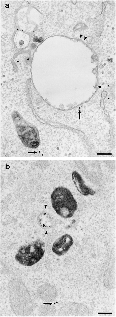
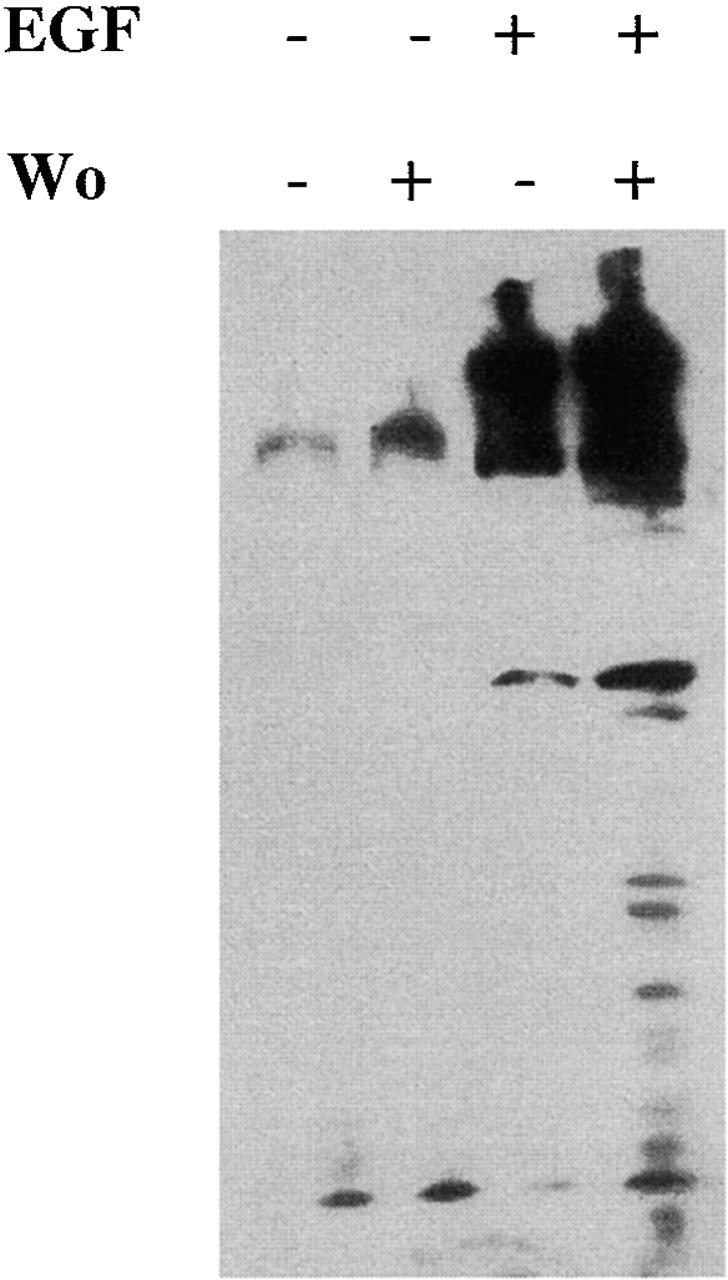
After internalization from the plasma membrane, activated EGF receptors (EGFRs) are delivered to multivesicular bodies (MVBs). Within MVBs, EGFRs are removed from the perimeter membrane to internal vesicles, thereby being sorted from transferrin receptors, which recycle back to the plasma membrane. The phosphatidylinositol (PI) 3'-kinase inhibitor, wortmannin, inhibits internal vesicle formation within MVBs and causes EGFRs to remain in clusters on the perimeter membrane. Microinjection of isotype-specific inhibitory antibodies demonstrates that the PI 3'-kinase required for internal vesicle formation is hVPS34. In the presence of wortmannin, EGFRs continue to be delivered to lysosomes, showing that their removal from the recycling pathway and their delivery to lysosomes does not depend on inward vesiculation. We showed previously that tyrosine kinase-negative EGFRs fail to accumulate on internal vesicles of MVBs but are recycled rather than delivered to lysosomes. Therefore, we conclude that selection of EGFRs for inclusion on internal vesicles requires tyrosine kinase but not PI 3'-kinase activity, whereas vesicle formation requires PI 3'-kinase activity. Finally, in wortmannin-treated cells there is increased EGF-stimulated tyrosine phosphorylation when EGFRs are retained on the perimeter membrane of MVBs. Therefore, we suggest that inward vesiculation is involved directly with attenuating signal transduction.


Figures

Figure 1.
Figure 1.
The effects of wortmannin on delivery of the EGFR to the lysosome. HEp-2 cells were incubated with HRP for 30 min at 37°C, chased for 3 h at 37°C, and then incubated with anti-EGFR gold and EGF for 1 h at 20°C, all in the absence of wortmannin. Cells were then chased for 1 h at 37°C in the absence (a) or presence of wortmannin (b and c). Note that in both the absence and the presence of wortmannin, the EGFR have, in many cases, reached the HRP-loaded lysosomes (arrows). In the presence of wortmannin, some MVBs (without HRP) are enlarged with comparatively few internal vesicles, and EGFRs on the perimeter membrane (asterisks) and some lysosomes (with HRP) are also enlarged (crosses). Bar, 0.2 μm.
Figure 2.
Figure 2.
The effects of wortmannin on EGF and EGFR degradation. (a) Cells were incubated with 125I-EGF for 1 h at 20°C, surface stripped, and chased at 37°C in the absence or presence of wortmannin (wo) for up to 2 h. Media samples were TCA precipitated to determine the percentage of degradation. (b) Cells were incubated with EGF for 1 h at 20°C and then chased at 37°C for up to 2 h in the absence or presence of wortmannin (wo). Cell lysates were analyzed by SDS-PAGE followed by Western blotting with an antibody against the cytoplasmic domain of the EGFR. Percentage degradation is calculated by comparison with the amount of EGFRs in cells not treated with EGF.
Figure 3.
Figure 3.
The effects of wortmannin on inward vesiculation in cells where the lysosomes have been cross-linked. HEp-2 cells were incubated with HRP for 30 min at 37°C, chased for 3 h at 37°C, and then incubated with DAB/H2O2 at 4°C to crosslink the lysosomes. Cells were then incubated with anti-EGFR gold and EGF at 20°C in the absence of wortmannin and then chased at 37°C in the absence (a) or the presence of wortmannin (b and c). Note that in the absence of wortmannin, MVBs with many internal vesicles and anti-EGFR gold predominantly on the internal vesicles accumulate (asterisks). In the presence of wortmannin, enlarged MVBs with very few internal vesicles accumulate (crosses), and the EGFR are clustered (arrowheads) on the perimeter membrane of the enlarged MVBs. L, lysosome. Bars: (a and b) 0.1 μm; (c) 0.5 μm.
Figure 4.
Figure 4.
The effects of wortmannin on the number of internal vesicles per MVB. HEp-2 cells were incubated with HRP for 30 min at 37°C, chased for 3 h at 37°C, and then incubated with DAB/H2O2 at 4°C to crosslink the lysosomes. Cells were then incubated with anti-EGFR gold and EGF at 20°C in the absence of wortmannin and then chased at 37°C in the absence or the presence of wortmannin. The total number of internal vesicles in 13 MVBs per treatment was estimated by analysis of 70-nm serial sections.
Figure 5.
Figure 5.
The effects of wortmannin on traffic of EGFRs and TRs through MVBs. HEp-2 cells were incubated with HRP for 30 min at 37°C, chased for 3 h at 37°C, and then incubated with DAB/H2O2 at 4°C to crosslink the lysosomes. Cells were then incubated with anti-EGFR gold (10 nm) and EGF for 1 h at 20°C in the absence of wortmannin and were then chased at 37°C for 1 h in the absence (a and b) or the presence of wortmannin (c). Cells were then permeabilized, fixed, and labeled with anti-TR antibody (5 nm gold). Note that in both the absence and the presence of wortmannin there are very few TRs on MVBs, but small vesicles labeling strongly for TRs (arrows) are often in close proximity. Bars, 0.1 μm.
Figure 6.
Figure 6.
The effects of microinjection with anti–PI 3-kinase antibodies on inward vesiculation in cells where the lysosomes have been cross-linked. HEp-2 cells were incubated with HRP for 30 min at 37°C, chased for 3 h at 37°C, and then incubated with DAB/H2O2 at 4°C to crosslink the lysosomes. Cells were then microinjected at 37°C with 20 nm BSA gold (arrows) alone (a) or coinjected with anti-hVPS34 antibody (b), anti-p110α antibody (c), or anti-p110β antibody (d). Cells were then incubated with 10 nm anti-EGFR gold (arrowheads) and EGF for 1 h at 20°C and were then chased at 37°C for 1 h. Microinjection with the anti-hVPS34 antibody caused the generation of enlarged MVBs containing few internal vesicles (asterisks). Microinjection with anti-p110α had no effect on MVB formation, whereas microinjection with p110β in some cases caused the generation of MVBs with comparatively few EGFRs, but they were not enlarged. Bars, 0.5 μm.
Figure 7.
Figure 7.
The effects of microinjection with anti–PI 3-kinase antibodies after incubation with EGF at 20°C. HEp-2 cells were incubated with HRP for 30 min at 37°C, chased for 3 h at 37°C, and then incubated with DAB/H2O2 at 4°C to crosslink the lysosomes. Cells were then incubated with 10 nm anti-EGFR gold (arrowheads) and EGF for 1 h at 20°C and were then microinjected with 20 nm BSA gold (arrows) and anti-hVPS34 (a) or anti-p110β (b) at 20°C before incubation at 20°C for a further 30 min and then chase at 37°C for 1 h. Microinjection of anti-hVPS34 caused the generation of enlarged MVBs with very few internal vesicles and EGFRs on the perimeter membrane. Microinjection of anti-p110β antibody caused the formation of small MVBs with few internal vesicles and few EGFRs. Bars, 0.2 μm.
Figure 8.
Figure 8.
The effects of wortmannin on EGF-stimulated tyrosine phosphorylation in cells were the lysosomes have been cross-linked. HEp-2 cells were incubated with HRP for 30 min at 37°C, chased for 3 h at 37°C, and then incubated with DAB/H2O2 at 4°C to crosslink the lysosomes. Cells were then incubated in the presence or absence of EGF at 20°C for 1 h and then chased at 37°C in the presence or absence of wortmannin (wo). Cell lysates were analyzed by SDS-PAGE followed by Western blotting with antiphosphotyrosine antibody.

References

    1. Barbieri, M.A., R.L. Roberts, A. Gumusboga, H. Highfield, C. Alvarez-Dominguez, A. Wells, and P.D. Stahl. 2000. Epidermal growth factor and membrane trafficking. EGF receptor activation of endocytosis requires rab5a. J. Cell Biol. 151:539–550. - PMC - PubMed
    1. Bright, N.A., B.J. Reaves, B.M. Mullock, and J.P. Luzio. 1997. Dense core lysosomes can fuse with late endosomes and are re-formed from the resultant hybrid organelles. J. Cell Sci. 110:2027–2040. - PubMed
    1. Bright, N.A., M.R. Lindsay, A. Stewart, and J.P. Luzio. 2001. The relationship between lumenal and limiting membranes in swollen late endocytic compartments formed after wortmannin treatment or sucrose accumulation. Traffic. 2:631–642. - PubMed
    1. Burke, B., K. Schooler, and H.S. Wiley. 2001. Regulation of epidermal growth factor receptor signaling by endocytosis and intracellular trafficking. Mol. Biol. Cell. 12:1897–1910. - PMC - PubMed
    1. Chin, L.-S., M.C. Raynor, X. Wei, H.-Q. Chen, and L. Li. 2001. Hrs interacts with sorting nexin 1 and regulates degradation of epidermal growth factor receptor. J. Biol. Chem. 276:7069–7078. - PubMed

Publication types

MeSH terms