. 2001 Dec 24;155(7):1307-18.
doi: 10.1083/jcb.200102017. Epub 2001 Dec 24.

Ser364 of connexin43 and the upregulation of gap junction assembly by cAMP

Affiliations

Ser364 of connexin43 and the upregulation of gap junction assembly by cAMP

E M TenBroek et al. J Cell Biol. .

Abstract

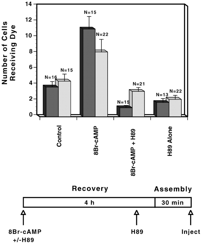
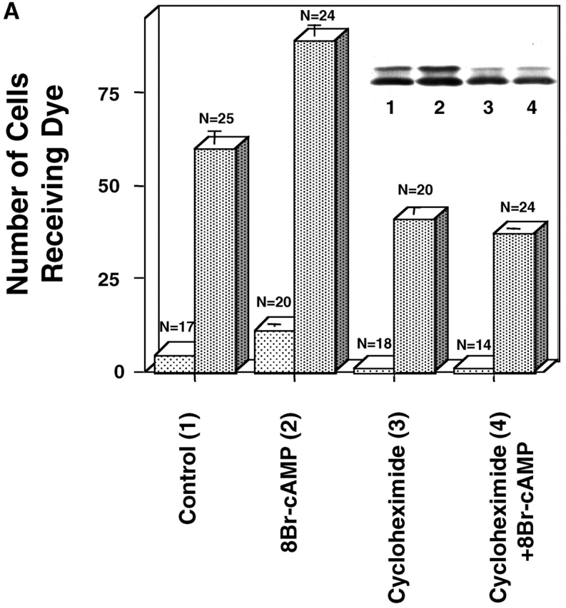
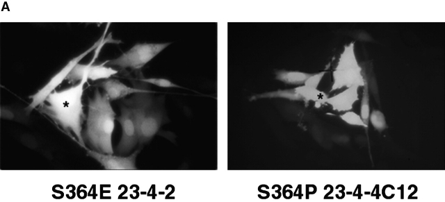
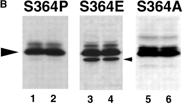
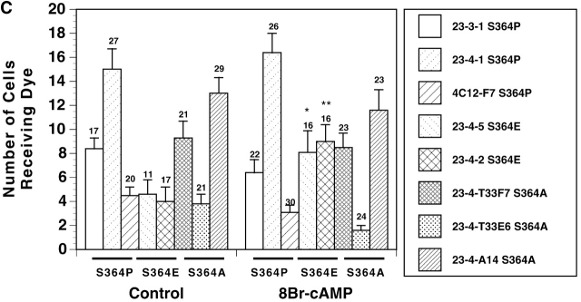
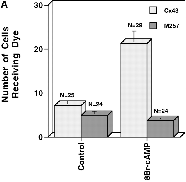
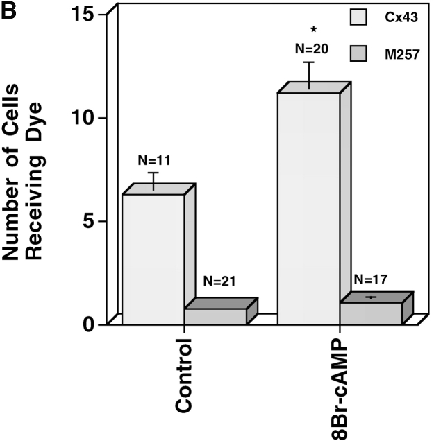
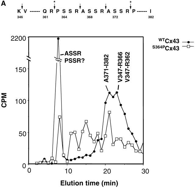
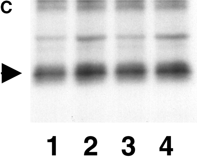
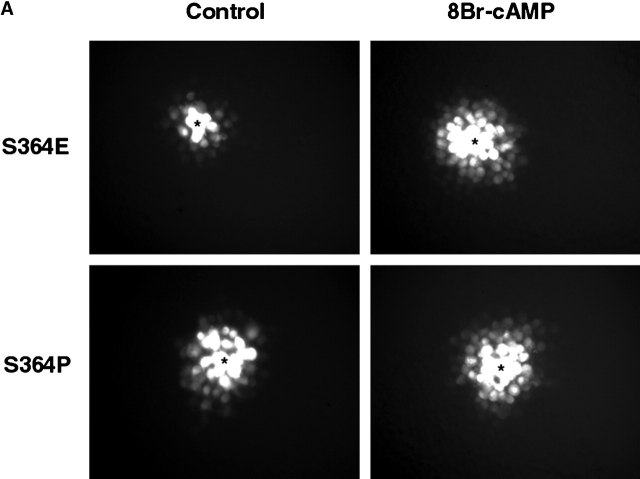
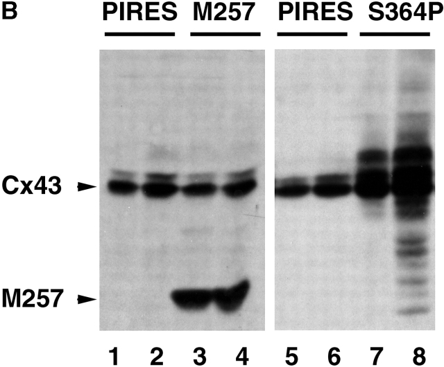
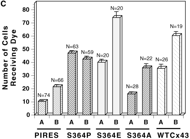
The assembly of gap junctions (GJs) is a process coordinated by growth factors, kinases, and other signaling molecules. GJ assembly can be enhanced via the elevation of cAMP and subsequent stimulation of connexon trafficking to the plasma membrane. To study the positive regulation of GJ assembly, fibroblasts derived from connexin (Cx)43 knockout (KO) and wild-type (WT) mice were transfected with WT Cx43 (WTCx43) or mutant Cx43. GJ assembly between untransfected WT fibroblasts or stably transfected WTCx43/KO fibroblasts was increased two- to fivefold by 8Br-cAMP, and this increase could be blocked by inhibition of cAMP-dependent protein kinase (PKA) or truncation of the Cx43 COOH terminus (CT). Although serine 364 (S364) of the Cx43 CT was determined to be a major site of phosphorylation, the molar ratio of Cx43 phosphorylation was not increased by 8Br-cAMP. Importantly, GJ assembly between either S364ECx43/KO or S364ECx43/WT fibroblasts was stimulated by 8Br-cAMP, but that between S364ACx43/KO or S364PCx43/KO fibroblasts was not stimulated, indicating that phosphorylation or a negative charge at S364 is required for enhancement of GJ assembly by cAMP. Furthermore, GJ assembly between S364ACx43/WT fibroblasts could be stimulated by 8Br-cAMP, but could not be between S364PCx43/WT fibroblasts. Thus, S364PCx43 interferes with enhanced GJ assembly when coexpressed with WTCx43.


Figures

Figure 1.
Figure 1.
Stimulation of GJ assembly after the elevation of cAMP. Two separate, representative experiments (dark vs. light bars) comparing GJ assembly between untreated WTCx43/KO fibroblasts (clone 22C-3) and cells treated with 8Br-cAMP, with both 8Br-cAMP and H89, or with H89 alone. (Dark bars) When present, H89 was added at recovery. (Light bars) When present, H89 was added 15 min before reaggregation.
Figure 2.
Figure 2.
The effects of cycloheximide on GJ assembly. Representative experiments comparing GJ assembly between WT fibroblasts (A) (clone 10-3) untreated (1) or treated with 8Br-cAMP (2), cycloheximide (3), or both cycloheximide and 8Br-cAMP (4). Cells were recovered for 4 h 23 min in suspension and reaggregated for 7 min (light bars) or recovered for 3.5 h in suspension and reaggregated for 1 h (dark bars). (Inset) Western immunoblot of WT fibroblasts after 1-h assembly (dark bars). (B) Immunolocalization of Cx43 using Cx43IF1 mouse monoclonal antibody (1:10), detected by goat anti–mouse Alexafluor 568 (1:500). WT fibroblasts were recovered for 3.5 h in suspension and reaggregated for 1 h before fixation. When present, 8Br-cAMP and/or cycloheximide were added at the beginning of recovery. (A) control; (B) 8Br-cAMP; (C) cycloheximide; (D) cycloheximide + 8Br-cAMP.
Figure 2.
Figure 2.
The effects of cycloheximide on GJ assembly. Representative experiments comparing GJ assembly between WT fibroblasts (A) (clone 10-3) untreated (1) or treated with 8Br-cAMP (2), cycloheximide (3), or both cycloheximide and 8Br-cAMP (4). Cells were recovered for 4 h 23 min in suspension and reaggregated for 7 min (light bars) or recovered for 3.5 h in suspension and reaggregated for 1 h (dark bars). (Inset) Western immunoblot of WT fibroblasts after 1-h assembly (dark bars). (B) Immunolocalization of Cx43 using Cx43IF1 mouse monoclonal antibody (1:10), detected by goat anti–mouse Alexafluor 568 (1:500). WT fibroblasts were recovered for 3.5 h in suspension and reaggregated for 1 h before fixation. When present, 8Br-cAMP and/or cycloheximide were added at the beginning of recovery. (A) control; (B) 8Br-cAMP; (C) cycloheximide; (D) cycloheximide + 8Br-cAMP.
Figure 5.
Figure 5.
GJ assembly between S364PCx43, S364ECx43 and S364ACx43/KO fibroblasts. (A) Example of Lucifer yellow dye transfer between S364PCx43 and S364ECx43/KO fibroblasts assembled overnight into confluent monolayers (*, injected cell). (B) Representative immunoblot of S364PCx43, S364ECx43, or S364ACx43/KO homogenates after GJ assembly and treatment without (1, 3, 5) or with (2, 4, 6) 8Br-cAMP for the entire assay. To detect S364PCx43 and S364ECx43 (arrowhead), membranes were labeled with a Cx43 antibody (1:8,000; Sigma-Aldrich) and detected with peroxi- dase-conjugated secondary antibody (1:10,000). A cross-reactive product, which was not consistently detected, is indicated below NP S364ECx43 (small arrowhead). (C) Compilation of representative GJ assembly experiments from S364PCx43, S364ECx43, and S364ACx43/KO fibroblasts. Cells were recovered for 4 h, reaggregated for 30 min, and, where indicated, treated with 8Br-cAMP for the entire 4.5 h of the assembly experiment. N values are above the error bars. *0.05 < P < 0.1; **0.01 < P < 0.02.
Figure 5.
Figure 5.
GJ assembly between S364PCx43, S364ECx43 and S364ACx43/KO fibroblasts. (A) Example of Lucifer yellow dye transfer between S364PCx43 and S364ECx43/KO fibroblasts assembled overnight into confluent monolayers (*, injected cell). (B) Representative immunoblot of S364PCx43, S364ECx43, or S364ACx43/KO homogenates after GJ assembly and treatment without (1, 3, 5) or with (2, 4, 6) 8Br-cAMP for the entire assay. To detect S364PCx43 and S364ECx43 (arrowhead), membranes were labeled with a Cx43 antibody (1:8,000; Sigma-Aldrich) and detected with peroxi- dase-conjugated secondary antibody (1:10,000). A cross-reactive product, which was not consistently detected, is indicated below NP S364ECx43 (small arrowhead). (C) Compilation of representative GJ assembly experiments from S364PCx43, S364ECx43, and S364ACx43/KO fibroblasts. Cells were recovered for 4 h, reaggregated for 30 min, and, where indicated, treated with 8Br-cAMP for the entire 4.5 h of the assembly experiment. N values are above the error bars. *0.05 < P < 0.1; **0.01 < P < 0.02.
Figure 3.
Figure 3.
Comparison of cells expressing WTCx43 or M257Cx43. (A) A representative experiment in which established monolayers of WTCx43/N2A and M257Cx43/N2A cells were treated with or without 8Br-cAMP for 4.5 h and coupling measured by microinjection of Lucifer yellow dye. (B) A representative experiment comparing de novo GJ assembly between WTCx43/N2A and M257Cx43/N2A cells treated for the entire experiment with or without 8Br-cAMP. *0.001 < P < 0.01
Figure 3.
Figure 3.
Comparison of cells expressing WTCx43 or M257Cx43. (A) A representative experiment in which established monolayers of WTCx43/N2A and M257Cx43/N2A cells were treated with or without 8Br-cAMP for 4.5 h and coupling measured by microinjection of Lucifer yellow dye. (B) A representative experiment comparing de novo GJ assembly between WTCx43/N2A and M257Cx43/N2A cells treated for the entire experiment with or without 8Br-cAMP. *0.001 < P < 0.01
Figure 4.
Figure 4.
S364 of Cx43 is phosphorylated in unstimulated WT Cx43/KO fibroblasts. (A) HPLC elution profile for 32P-labeled Cx43 peptides isolated from cells expressing WTCx43 (•) or S364PCx43 (□). The elution position of certain peptides is indicated. Amino acids 346–382 of Cx43 and tryptic cleavage sites are depicted above the profile (arrows). Asterisks denote sites that may only be partially cleaved by trypsin. (B) Autoradiogram depicting in vitro phosphorylation of Cx GST fusion proteins. (Lane 1) Purified GST CTCx56 phosphorylated by PKA. (Lane 2) Purified GST CTCx43 phosphorylated by of PKA. (Lane 3) Purified GST CTCx43 phosphorylated by PKC. (C) Phosphorylation of Cx43 after treatment with 8Br-cAMP. Autoradiogram showing results of two separate experiments in which confluent monolayers of WT fibroblasts (10-3) were loaded with radiolabeled [32P]i in the presence or absence of 8Br-cAMP (Materials and methods). (1 and 3) Untreated. (2 and 4) Treated with 8Br-cAMP.
Figure 4.
Figure 4.
S364 of Cx43 is phosphorylated in unstimulated WT Cx43/KO fibroblasts. (A) HPLC elution profile for 32P-labeled Cx43 peptides isolated from cells expressing WTCx43 (•) or S364PCx43 (□). The elution position of certain peptides is indicated. Amino acids 346–382 of Cx43 and tryptic cleavage sites are depicted above the profile (arrows). Asterisks denote sites that may only be partially cleaved by trypsin. (B) Autoradiogram depicting in vitro phosphorylation of Cx GST fusion proteins. (Lane 1) Purified GST CTCx56 phosphorylated by PKA. (Lane 2) Purified GST CTCx43 phosphorylated by of PKA. (Lane 3) Purified GST CTCx43 phosphorylated by PKC. (C) Phosphorylation of Cx43 after treatment with 8Br-cAMP. Autoradiogram showing results of two separate experiments in which confluent monolayers of WT fibroblasts (10-3) were loaded with radiolabeled [32P]i in the presence or absence of 8Br-cAMP (Materials and methods). (1 and 3) Untreated. (2 and 4) Treated with 8Br-cAMP.
Figure 6.
Figure 6.
GJ assembly following the expression of vector, S364PCx43, S364ECx43, S364ACx43, or WTCx43 in WT fibroblasts. (A) Comparison of GJ assembly between WT fibroblasts (10-3) transfected with either S364PCx43 or S364ECx43 and treated with or without 8Br-cAMP for the entire experiment (*, injected cell). (B) Example of immunoblots of cell homogenates following two different assembly experiments using either WT fibroblasts transiently transfected with M257Cx43 or S364PCx43. Cells were transfected 48 h prior to experimentation with either vector alone (pIRES; 1, 2, 5, and 6) or mutant construct (3, 4, 7, and 8). Cells were dissociated and recovered for 4 h 15 or 23 min, reaggregated for 15 (M257Cx43) or 7 (S364PCx43) min, and treated either without (1 and 3) or with (2 and 4) 8Br-cAMP for the entire GJ assembly experiment. To detect endogenous WTCx43 and transfected M257Cx43 (arrowhead, first panel) membranes were labeled with NH2-terminal Cx43 antibody (1:8,000) detected with goat anti–rabbit peroxidase antibody (1:10,000). To detect mutant and WTCx43 expression (second panel), membranes were labeled with Cx43 CT antibody (1:8,000; Sigma-Aldrich) detected with goat anti–rabbit peroxidase antibody (1:10,000). (C) Compilation of trials in which WT fibroblasts were transfected with either vector alone, mutant, or WT Cx43 and GJ assembly monitored without (A) or with (B) 8Br-cAMP.
Figure 6.
Figure 6.
GJ assembly following the expression of vector, S364PCx43, S364ECx43, S364ACx43, or WTCx43 in WT fibroblasts. (A) Comparison of GJ assembly between WT fibroblasts (10-3) transfected with either S364PCx43 or S364ECx43 and treated with or without 8Br-cAMP for the entire experiment (*, injected cell). (B) Example of immunoblots of cell homogenates following two different assembly experiments using either WT fibroblasts transiently transfected with M257Cx43 or S364PCx43. Cells were transfected 48 h prior to experimentation with either vector alone (pIRES; 1, 2, 5, and 6) or mutant construct (3, 4, 7, and 8). Cells were dissociated and recovered for 4 h 15 or 23 min, reaggregated for 15 (M257Cx43) or 7 (S364PCx43) min, and treated either without (1 and 3) or with (2 and 4) 8Br-cAMP for the entire GJ assembly experiment. To detect endogenous WTCx43 and transfected M257Cx43 (arrowhead, first panel) membranes were labeled with NH2-terminal Cx43 antibody (1:8,000) detected with goat anti–rabbit peroxidase antibody (1:10,000). To detect mutant and WTCx43 expression (second panel), membranes were labeled with Cx43 CT antibody (1:8,000; Sigma-Aldrich) detected with goat anti–rabbit peroxidase antibody (1:10,000). (C) Compilation of trials in which WT fibroblasts were transfected with either vector alone, mutant, or WT Cx43 and GJ assembly monitored without (A) or with (B) 8Br-cAMP.

Similar articles

Cited by

References

    1. Atkinson, M.M., P.D. Lampe, H.H. Lin, R. Kollander, X.R. Li, and D.T. Kiang. 1995. Cyclic AMP modifies the cellular distribution of connexin43 and induces a persistent increase in the junctional permeability of mouse mammary tumor cells. J. Cell Sci. 108:3079–3090. - PubMed
    1. Beardslee, M.A., J.G. Laing, E.C. Beyer, and J.E. Saffitz. 1998. Rapid turnover of connexin43 in the adult rat heart. Circ. Res. 83:629–635. - PubMed
    1. Berthoud, V.M., M.B. Rook, O. Traub, E.L. Hertzberg, and J.C. Saez. 1993. On the mechanisms of cell uncoupling induced by a tumor promoter phorbol ester in clone 9 cells, a rat liver epithelial cell line. Eur. J. Cell Biol. 62:384–396. - PubMed
    1. Berthoud, V.M., E.C. Beyer, W.E. Kurata, A.F. Lau, and P.D. Lampe. 1997. The gap-junction protein connexin 56 is phosphorylated in the intracellular loop and the carboxy-terminal region. Eur. J. Biochem. 244:89–97. - PubMed
    1. Beyer, E.C., D.L. Paul, and D.A. Goodenough. 1987. Connexin43: a protein from rat heart homologous to a gap junction protein from liver. J. Cell Biol. 105:2621–2629. - PMC - PubMed

Publication types