. 2002 Jan 1;22(1):1-9.
doi: 10.1523/JNEUROSCI.22-01-00001.2002.

Protein kinase modulation of a neuronal cation channel requires protein-protein interactions mediated by an Src homology 3 domain

Affiliations

Protein kinase modulation of a neuronal cation channel requires protein-protein interactions mediated by an Src homology 3 domain

Neil S Magoski et al. J Neurosci. .

Abstract

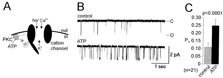
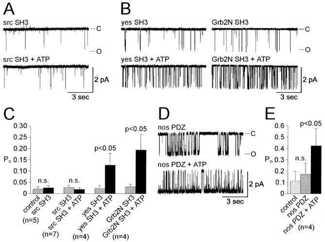
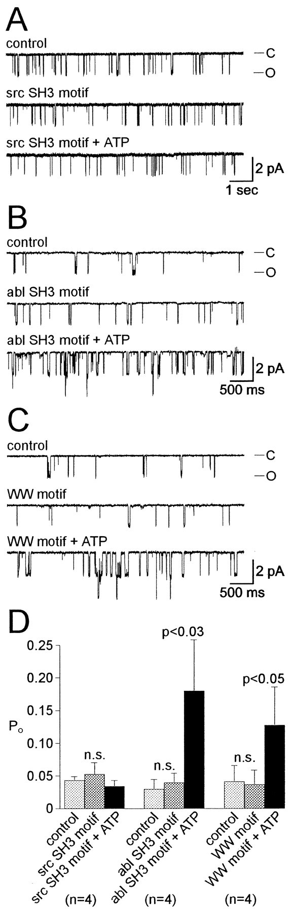
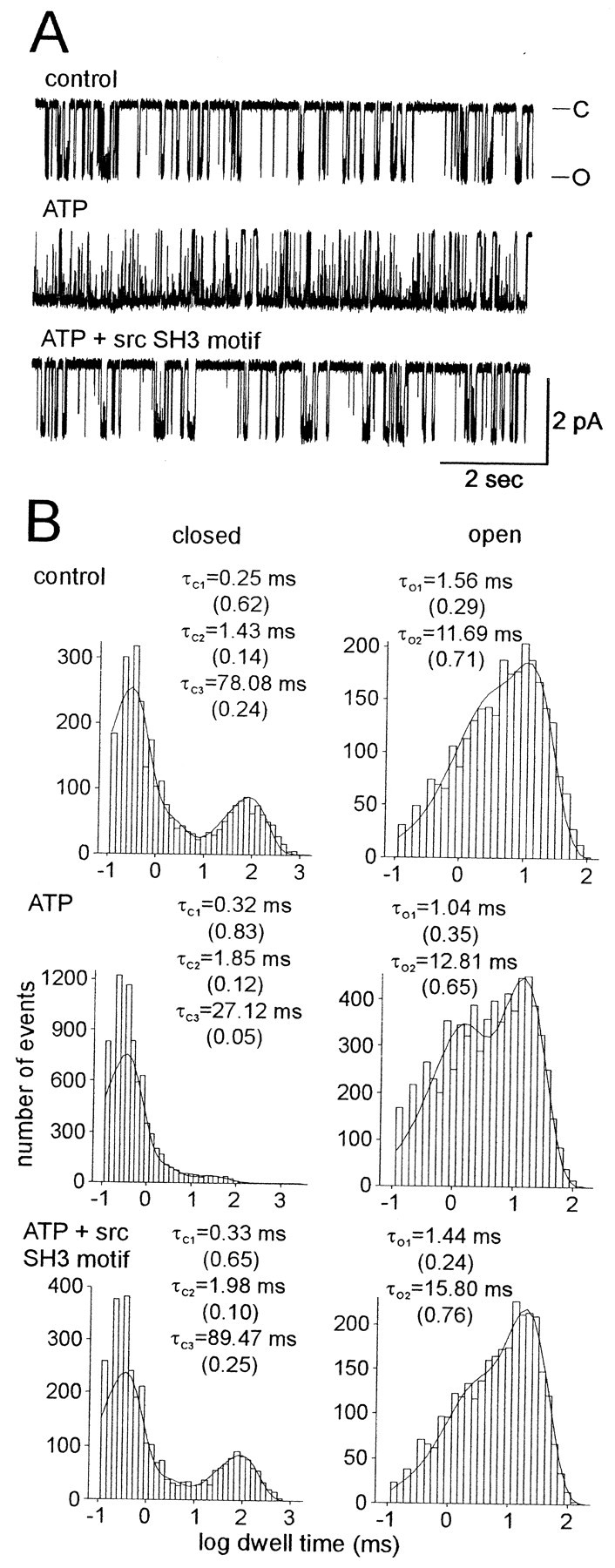
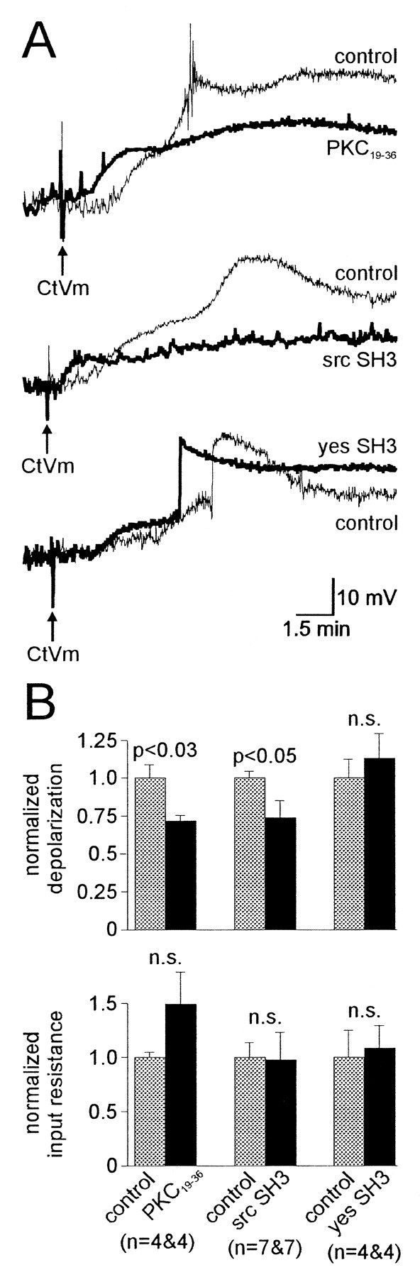
Accumulating evidence suggests that many ion channels reside within a multiprotein complex that contains kinases and other signaling molecules. The role of the adaptor proteins that physically link these complexes together for the purposes of ion channel modulation, however, has been little explored. Here, we examine the protein-protein interactions required for regulation of an Aplysia bag cell neuron cation channel by a closely associated protein kinase C (PKC). In inside-out patches, the PKC-dependent enhancement of cation channel open probability could be prevented by the src homology 3 (SH3) domain, presumably by disrupting a link between the channel and the kinase. SH3 and PDZ domains from other proteins were ineffective. Modulation was also prevented by an SH3 motif peptide that preferentially binds the SH3 domain of src. Furthermore, whole-cell depolarizations elicited by cation channel activation were decreased by the src SH3 domain. These data suggest that the cation channel-PKC association may require SH3 domain-mediated interactions to bring about modulation, promote membrane depolarization, and initiate prolonged changes in bag cell neuron excitability. In general, protein-protein interactions between ion channels and protein kinases may be a prominent mechanism underlying neuromodulation.


Figures

Fig. 1.
Fig. 1.
ATP increases cation channelPO in excised, inside-out patches from cultured bag cell neurons. A, Model of the cation channel based on Wilson et al. (1998). Under physiological conditions, the channel passes Na+, K+, and Ca2+. ATP is used as a phosphate source in a phosphotransfer reaction catalyzed by a closely associated PKC-like enzyme (PKC). This phosphorylation of either the cation channel itself or a nearby protein results in enhanced activity. The purpose of the present study was to examine the nature of the interaction between the cation channel and the kinase.B, Top trace, At a holding potential of −60 mV, cation channel activity was observed as unitary inward current steps of ∼2 pA. For this and subsequent figures, the closed state (−C) is at the top, and the open state (−O) is at the bottom of thetrace. Bottom trace, Bath application of 1 mm ATP to the cytoplasmic face of the patch resulted in a marked and sustained increase in POfrom 0.012 to 0.093. The PO rose to its elevated level within 10 sec of adding ATP; most likely, diffusion of ATP from the site of bath application (pipetting location; see Materials and Methods) was the determining factor in this time course.C, Grouped data for control experiments used in the present study. The increase in cation channelPO produced by ATP was statistically significant. The PO was calculated over the entire time of recording before and then after ATP (typically 1–3 min in each condition; see Materials and Methods). The nvalue refers to the number of patches.
Fig. 2.
Fig. 2.
The ATP-induced increase in channelPO is blocked by the src SH3 protein–protein interaction domain. A, Application of a 100 nm concentration of the GST fusion protein of the SH3 domain from the tyrosine kinase src prevented the enhancement of cation channel PO by ATP (0.005 in src SH3 vs 0.005 in src SH3 plus 1 mm ATP). The domain was bath-applied to the cytoplasmic face of the patch, which in this case contained two channels, and was followed by ATP ∼2 min later. The patch was held at −60 mV. B, The block is specific to the src form of the SH3 domain, because GST-fused SH3 domains from the related tyrosine kinase yes (left traces) (PO = 0.014 in 375 nm yes SH3 vs 0.116 in yes SH3 plus 1 mm ATP) or the N-terminal SH3 domain from the grb2 adaptor protein (right traces;PO = 0.014 in 300 nm grb2N SH3 vs 0.116 in grb2N SH3 plus 1 mm ATP) fail to inhibit the ATP-induced PO increase. Both patches were held at −60 mV. C, Summary data for the effect of SH3 domains on the cation channel response to ATP. The src SH3 domain had no effect itself on channel PO; furthermore, it prevented ATP from enhancingPO. This is in contrast to the SH3 domains from yes or grb2N, neither of which impeded the significant elevation of PO by ATP. For C andE, the n values refer to the number of patches; n.s., Not significant. D, Exposure to the GST-fused PDZ interaction domain from the enzyme NOS did not prevent ATP from increasing cation channelPO (0.208 in 130 nm NOS PDZ vs 0.573 in NOS PDZ plus 1 mm ATP). The patch was held at −60 mV. E, Summary data for the effect of the NOS PDZ domain on cation channel activity and the ATP response. The PDZ domain did not alter cation channel activity, nor did it hinder ATP from significantly enhancing PO.
Fig. 3.
Fig. 3.
An src SH3 motif peptide blocks the ATP-induced increase in PO. A, In keeping with an SH3-like domain-mediated interaction being involved in the channel–kinase association, a 1 μm concentration of an SH3 motif peptide (NH2-LAS RPL PLL PNSAPGQ-COOH, src SH3 motif) that preferentially binds to the src SH3 domain blocked the ATP response (PO = 0.038 in controls vs 0.037 in src SH3 peptide vs 0.025 in src SH3 peptide plus 1 mm ATP). The src SH3 motif peptide was in the bath for ∼2 min before the addition of ATP. The patch was held at −60 mV. B, To determine the specificity of the disruption of the channel–kinase complex by the src SH3 motif, a second SH3 motif (NH2-SGSGSR PPRWS PPV PLPTSLDSR-COOH, abl SH3 motif) was used that preferentially binds the abl SH3 domain over the src SH3 domain. A 1 μm dose of abl SH3 motif peptide failed to block the ATP response (PO = 0.023 in controls vs 0.037 in abl SH3 peptide vs 0.073 in abl SH3 peptide plus 1 mm ATP). C, As an additional control for nonspecific effects, a 1 μm concentration of the WW motif peptide (NH2-LKLPDYWESSAS-COOH) was applied to the cytoplasmic face of a cation channel-containing patch. This peptide did not interfere with the ATP-induced increase inPO (0.042 in controls vs 0.036 in WW peptide vs 0.055 in WW peptide plus 1 mm ATP).D, Summary data for the effect of src SH3, abl SH3, and WW motif peptides on the cation channel response to ATP. Of the two SH3 motif peptides, neither had a significant impact on channel activity; however, the src SH3 motif peptide but not the abl SH3 motif peptide inhibited the ATP-induced POelevation. Furthermore, not only did the WW motif peptide have no effect on cation channel PO, it also failed to prevent an increase in PO by ATP. The n values refer to the number of patches;n.s., Not significant.
Fig. 4.
Fig. 4.
The src SH3 motif peptide reverses the ATP-induced increase in PO. A, A patch held at −60 mV displayed a single, rather active cation channel under control conditions (PO = 0.203). With the application of 1 mm ATP to the cytoplasmic face, the activity was markedly elevated (PO = 0.530). However, when 1 μm SH3 motif peptide was added along with the ATP, the activity of the channel fell back toward control levels (PO = 0.316).B, Given that this patch appeared to contain only a single cation channel, kinetic analysis was possible. The histograms represent single- channel closed and open dwell times, fit with a sum of exponentials by using the maximum likelihood estimator method and a simplex search (see Materials and Methods). The time constants of the exponentials are given in the inset of each graph, with the proportion or fractional contribution to the area under the curve of each exponential shown in brackets under its respective time constant. During the control period (top graphs), cation channel closed times were best described by three exponentials (τC1,τC2, andτC3) and the open times were best described by two exponentials (τO1and τO2). After exposure to ATP (middle graphs), the τC1 or τC2 showed little change; however, τC3 was substantially reduced both in duration (a 65% decrease, from ∼78 to ∼27 msec) and in the proportion of events that it described (a 80% decrease, from 0.25 to 0.05). For open times during ATP, there was a modest shift, from τO2 to τO1, in the proportion of events described. After application of the SH3 motif peptide (bottom graphs), τC3 values returned to near control levels. Similarly, the number of events described by τO1 and τO2 shifted back toward control levels.
Fig. 5.
Fig. 5.
CtVm-induced depolarization is reduced by both a PKC inhibitor and the src SH3 domain. A, Whole-cell current-clamp recordings of depolarizations elicited by CtVm (100 μg/ml) in cultured bag cell neurons. CtVm responses in neurons dialyzed with control intracellular saline (gray line) are displayed along with responses of neurons dialyzed with intracellular saline containing 50 μmPKC19–36, 300 nm src SH3 domain, or 300 nm yes SH3 domain (black line). Neurons were dialyzed for 45 min before CtVm bath application (arrow). The peak CtVm- induced depolarization for neurons dialyzed with either PKC19–36 (top traces) or src SH3 domain (middle traces) was reduced compared with controls; however, dialysis with yes SH3 domain (bottom traces) did not have a obvious impact on the peak depolarization. The control CtVm response depicted in the top traces shows some action potentials approximately halfway through the recording period; the spikes are truncated because of the low digitization rate and the display scale. Differences in the onset of the depolarization are not significant and are most likely attributable to experimental variability. B, Summary data for the effect of PKC19–36, src SH3 domain, or yes SH3 domain on the bag cell neuron membrane potential response to CtVm and on input resistance before CtVm application. Top graph, On average, both PKC19–36 and src SH3 domain dialysis significantly reduced the depolarization by approximately one-third compared with controls. The SH3 domain from yes did not produce any significant change in the CtVm-induced depolarization. Bottom graph, Input resistance (see Materials and Methods) was measured at the end of dialysis and just before the application of CtVm. Compared with controls, none of the agents introduced into the neurons had a significant effect on input resistance. The apparent but nonsignificant elevation after PKC19–36 introduction was attributable to a single neuron showing an inexplicable increase in input resistance over the dialysis period. The labels apply to both thetop and bottom graphs, and then values refer to the number of control and experimental neurons examined in a given set. n.s., Not significant.

Similar articles

Cited by

References

    1. Alonso A, Llinas RR. Subthreshold Na+-dependent theta-like rhythmicity in stellate cells of entorhinal cortex layer II. Nature. 1989;342:175–177. - PubMed
    1. Bielefeldt K, Jackson MB. Phosphorylation and dephosphorylation modulate a Ca2+-activated K+ channel in rat peptidergic nerve terminals. J Physiol (Lond) 1994;475:241–254. - PMC - PubMed
    1. Cantrell AR, Tibbs VC, Westenbrock RE, Scheuer T, Catterall WA. Dopaminergic modulation of voltage-gated Na+ current in rat hippocampal neurons requires anchoring of cAMP-dependent protein kinase. J Neurosci 19 2000. RC21:1–6. - PMC - PubMed
    1. Chung S, Reinhart PH, Martin BL, Brautigan D, Levitan IB. Protein kinase activity closely associated with a reconstituted calcium-activated potassium channel. Science. 1991;253:560–562. - PubMed
    1. Coghlan VM, Perrino BA, Howard M, Langeberg LK, Hicks JB, Gallatin WM, Scott JD. Association of protein kinase A and protein phosphatase 2B with a common anchoring protein. Science. 1995;267:108–111. - PubMed

Publication types

MeSH terms

LinkOut - more resources